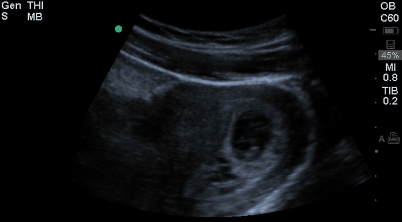
The team evaluated a 21 yo F with nausea and emesis and used bedside US to evaluate the known pregnancy. Take a look at the unlabeled still image (Image 1).
Image 1

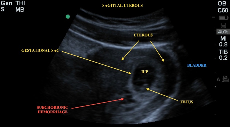
This is a sagittal section through the uterus using the curvilinear probe with the indicator towards the head of the patient and the probe placed midline in the suprapubic region. Do you notice anything in addition to the intrauterine pregnancy (IUP)? Note the hypoechoic area adjacent to the gestational sac (Image 2). This represents a subchorionic hemorrhage. In this type of hemorrhage, blood collects between the uterine wall and the chorionic membrane. The clinical significance of a small, incidental subchorionic hemorrhage is debated but is generally followed as an outpatient. Enlarging symptomatic hematomas, hematomas as a result of abdominal trauma, and hematomas found outside the first trimester may need more urgent evaluation by OB.
Image 2

In a 2003 article by Nagy et al., intrauterine hematomas were found in 3.1% of the study population (N=6488). Authors concluded that the presence of an intrauterine hematoma may identify a population at increased risk for adverse pregnancy outcome - to include miscarriage, preeclampsia, abruptio placentae, and preterm labor.
Recall that when using bedside US to evaluate asymptomatic pt with a +BHCG or +UPT, our goal as emergency providers is to find an IUP, not an ectopic. The primary question in this type of patient is "Is there an intrauterine pregnancy?" Recall that we define IUP in the ED as a: 1) Gestational Sac with a 2) Fetal Pole or Yolk Sac (or both), 3) Surrounded by a thick rim of the myometrium (myometrial mantle of 8 mm or more in two planes). If the answer to this question is no, then there is an ECTOPIC PREGNANCY until proven otherwise.
Date: July 2012
Image credit: Drs. Deepa Patel and Richard Wang.

