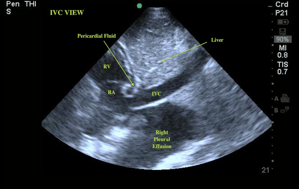
Normally we image the IVC to give us a ballpark assessment of a patient’s volume status, and preload to the right heart. This image, however, provides us with some additional valuable information. Notice the IVC transversing the liver and draining into the right atrium. An additional small amount of fluid can be seen surrounding the heart. This is a trace pericardial effusion. The IVC view shows us the most dependent portion of the pericardial sac and is the most sensitive for picking up a small pericardial effusion. In this case, this amount of fluid is unlikely to be clinically significant. We can also see a second large fluid collection in the far field of the image. This is a large right pleural effusion.

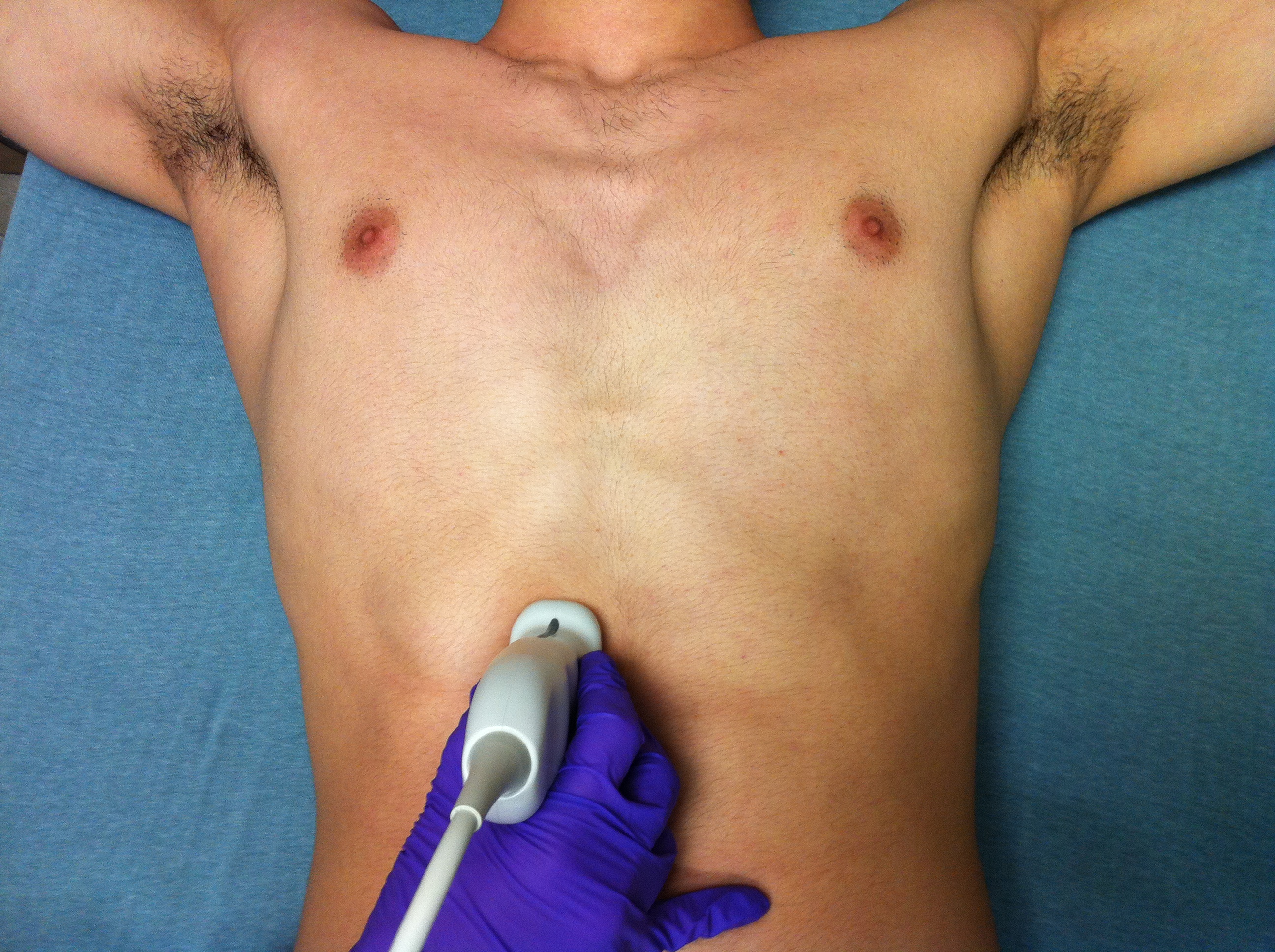
To get this view place the curvilinear probe subxiphoid, with the indicator pointed towards the head. Slide the probe towards the patient's right and angle slightly towards the heart. This may require some adjustments (fanning the probe left or right, angling more into the chest, or more towards the abdomen) to get a perfect view. (See the second image) The IVC can sometimes be confused with the aorta or the gallbladder, so it is important to confirm that what you are looking at is truly the IVC.

Date: January 2012
Author: Dr. Lemos

