The IOW comes from evaluating a 95-yo-female with shortness of breath and peripheral edema, the following video was obtained:
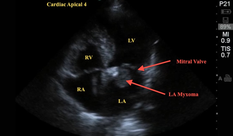
Can you identify the cardiac view and abnormality? See the labeled still image - IOW Still A4V.
Image 1: IOW Still A4V
 .
.
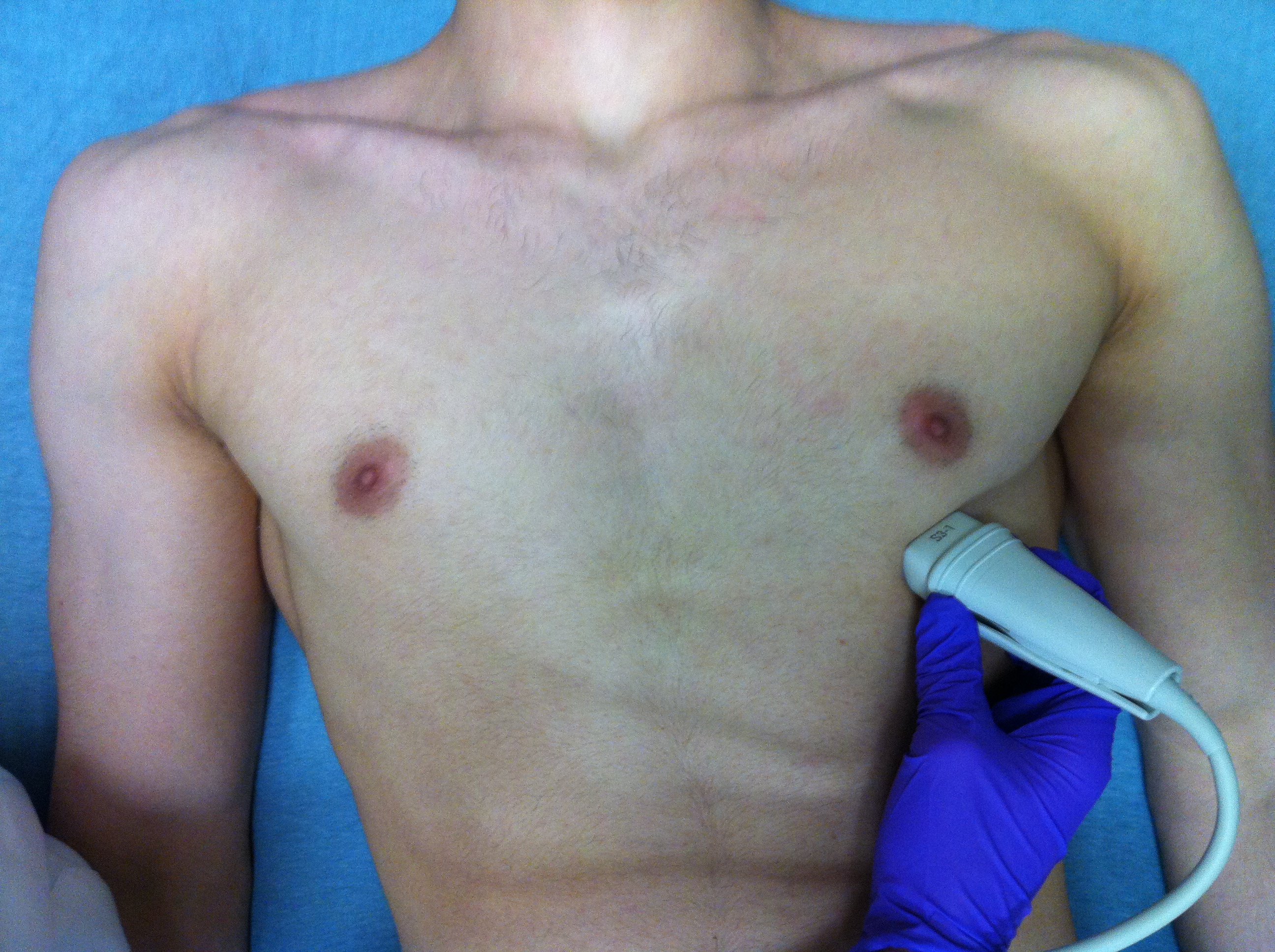
This is an apical 4-chamber view (A4C) of the heart obtained by placing the cardiac probe at the point of maximal impulse (PMI) and looking up into the chest, with the indicator to the RIGHT of the patient (see Image 2).
Image 2

This is an excellent view to permit comparison of RIGHT and LEFT sides of the heart and also allow a view of global wall motion.
Note how this video clip captures a mobile mass - adherent to the wall of the left atrium (LA) and swinging toward the left ventricle (LV).
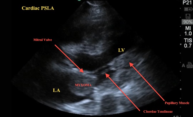
In another view, the parasternal long-axis (PSLA), see how the mass moves but remains in the LA (see labeled still image, IOW Still PSLA).
Image 3: IOW Still PSLA

The patient was known to have an atrial myxoma and despite medication, compliance remained increasingly symptomatic. Atrial myxomas are primary cardiac tumors and VERY rare - when found, most (75%) reside in the LA. As these enlarge they alter cardiac blood flow, can mimic mitral stenosis, and may result in a variety of signs/symptoms including SOB, dyspnea, pulmonary and peripheral edema, dizziness, and dysrhythmias. Thromboembolism and peripheral emboli may also occur. Surgical removal and mitral valve repair are generally the treatment of choice.
Date: 2012

