This week’s image is an elderly male who presented with persistent periumbilical abdominal pain. A Point-of-Care ultrasound of the abdomen was performed and the following images were obtained:
Image 1
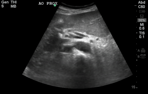
Image 2
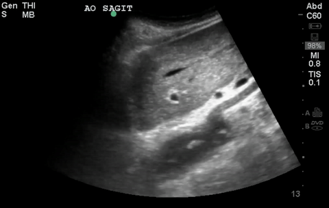
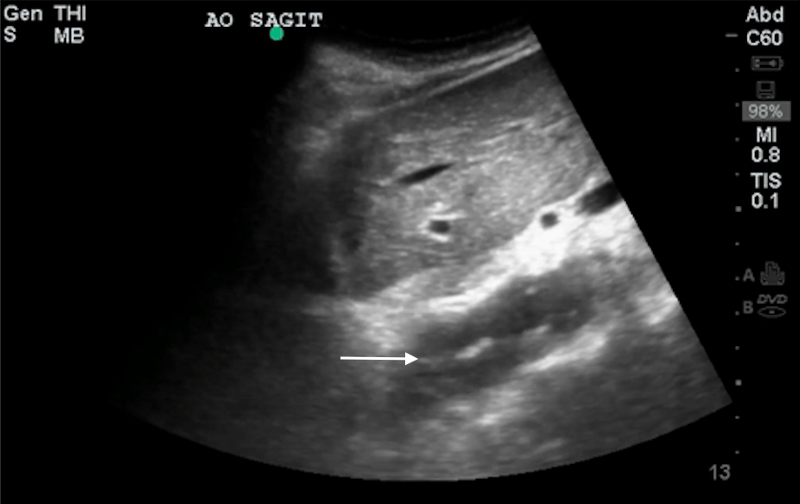
These images contain some key findings. As you can see on the short axis view of the aorta, it appears to be quite tortuous. A dissection flap is noted (white arrow) both in the long and short axis view of the vessel as labeled in the still images below.
Image 3
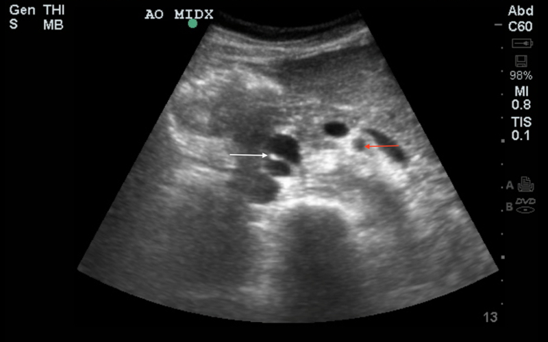
Image 4
Note that not all patients will have the textbook anatomy. For instance, this patient’s aorta is quite displaced in its relationship with the superior mesenteric artery (red arrow) due to its tortuosity in the short axis image provided. The ability to mentally visualize and identify structures on ultrasound is of paramount importance in accurate imaging and diagnostics. If you are lost, try to orient to structures you can confidently identify with consistent anatomy (such as the spine in this image) and use that landmark to orient to confusing anatomic structures. Focused ultrasonography can serve as a valuable tool in answering specific clinical questions based on history and physical examination.