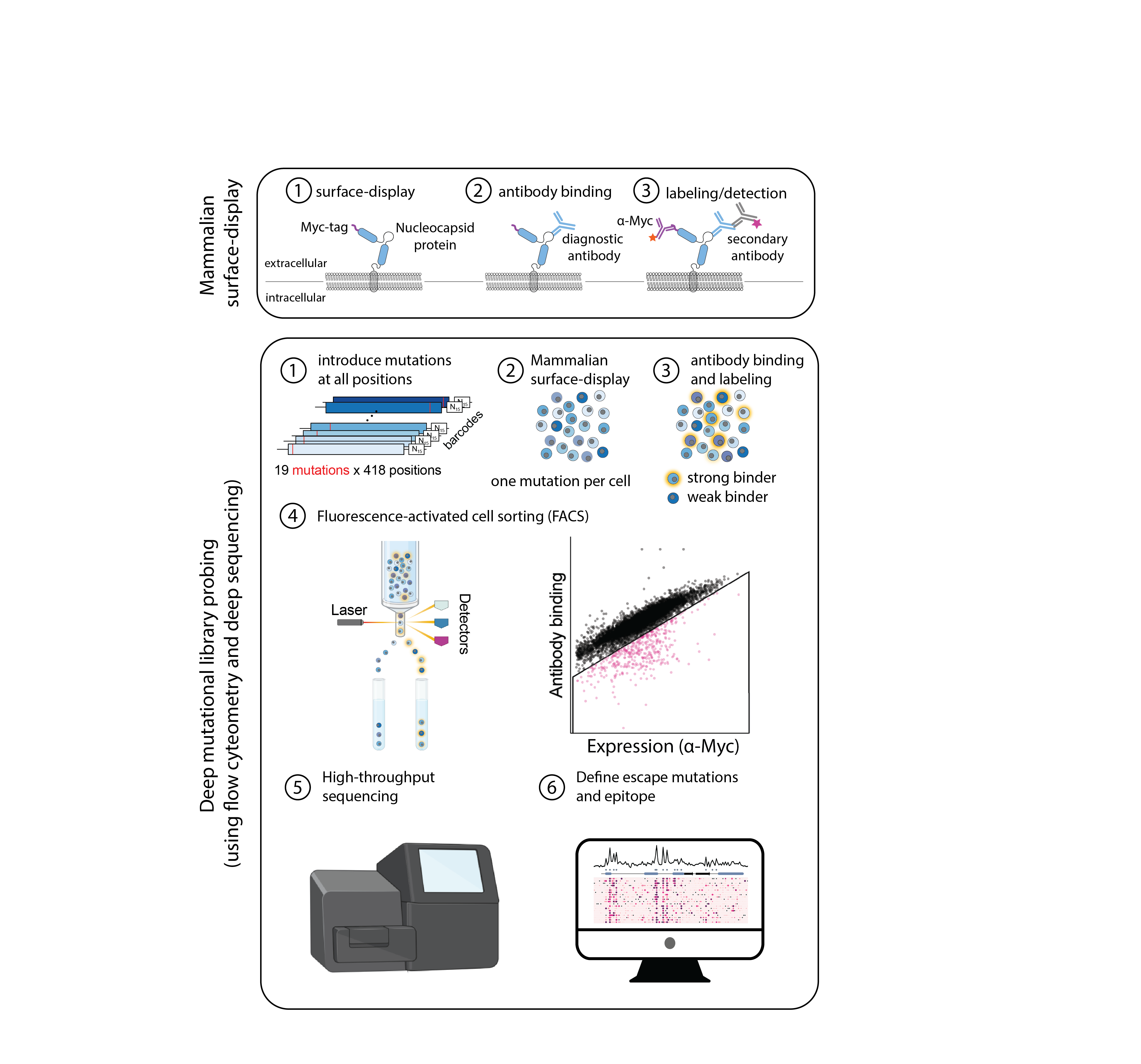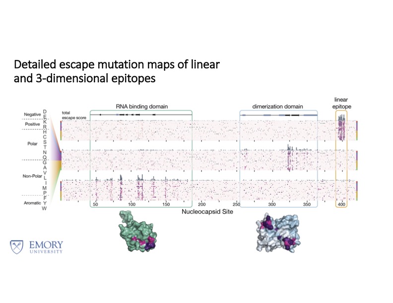
By combining mammalian surface display with high throughput deep mutational scanning (DMS), it is possible to map the epitopes of stably-folded, full length proteins. We achieve this by creating a variant library of our protein of interest, packaged inside a lentiviral expression construct, which includes green fluorescent protein (GFP) and a Myc-tag, to control for differences in surface expression. This allows for direct probing of antibody/ligand binding in a single, high throughput experiment via fluorescence activated cell sorting (FACS). Tighter and weaker binders can be separately sorted, and their sequences compared against an input, reference population, thus identifying mutations which enhance or diminish binding. This data can be represented on 2D heatmaps, where regions of pronounced decreased binding suggest direct disruption and so typically correspond to epitopes. Some epitopes are linear; others are conformational, and these become apparent if plotted on 3D structures.
KD values can be derived by running titrations, where the antibody/ligand is sorted against the protein of interest over a range of concentrations. Cell sorting is achieved via a binning strategy, and the shift in the relative population of bins allows for a binding curve to be plotted. These KD predictions can be validated by expressing selected mutants and analyzing with SPR/BLI.
This DMS technology has been used to map epitopes and predict the mutations which would cause the virus to escape antibody detection within commercially available rapid antigen tests for the SARS-CoV-2 nucleocapsid protein. Currently, we are expanding our analysis to cover various flu strain to develop next generation biologics.


