Course Overview
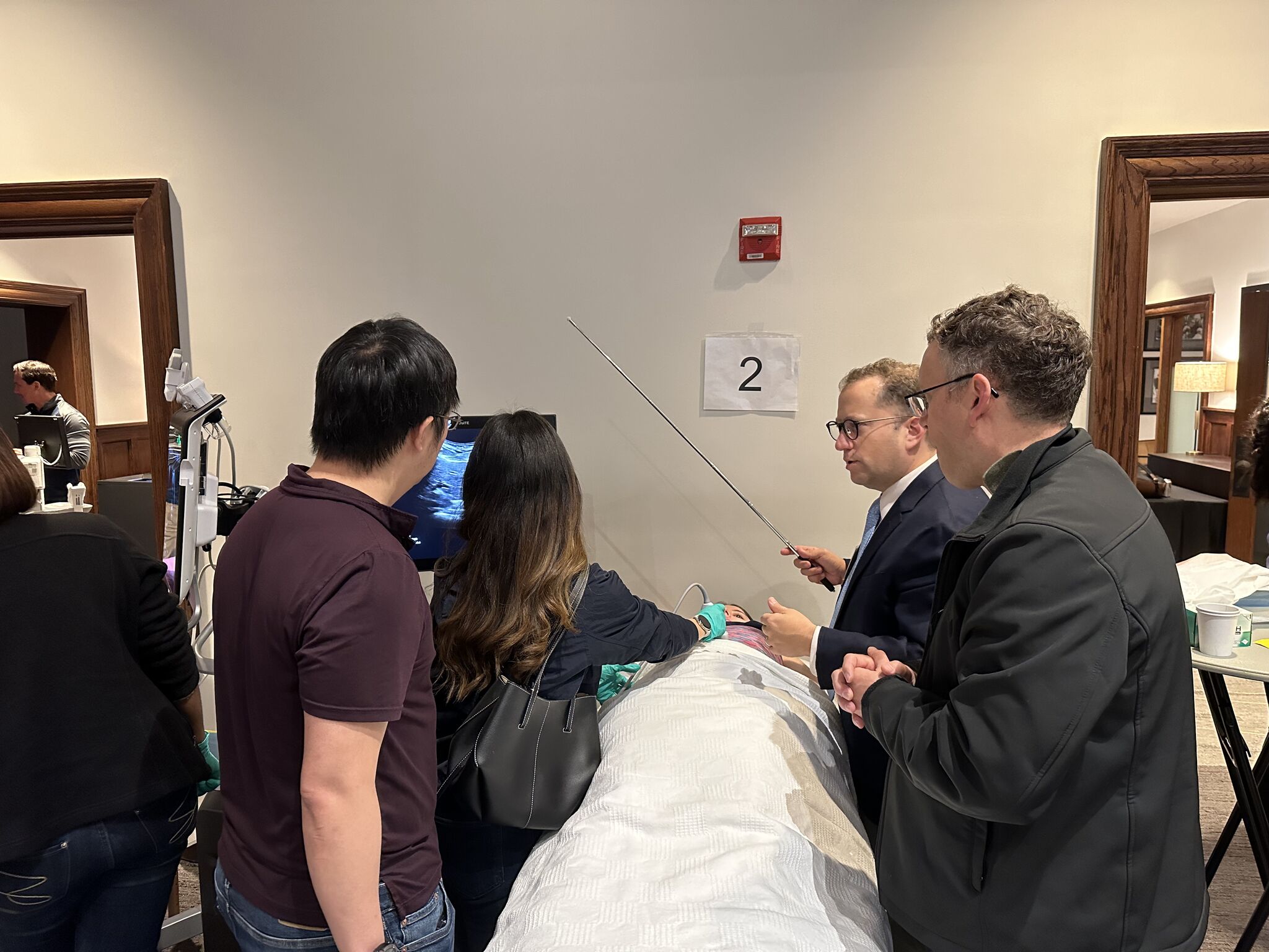
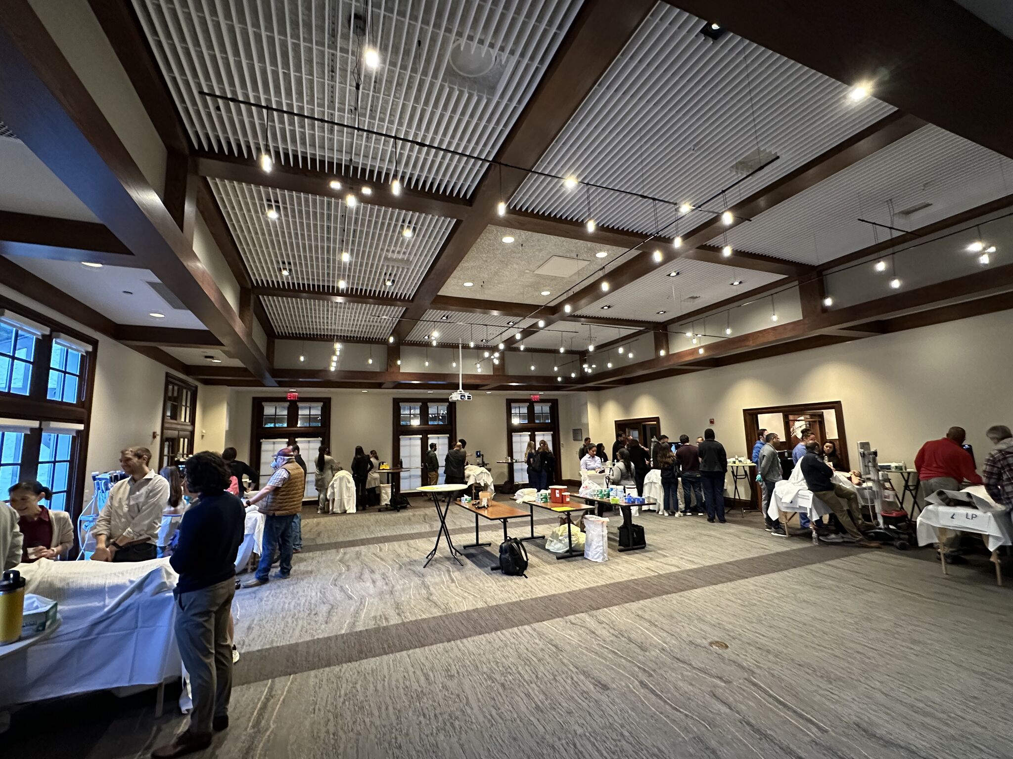
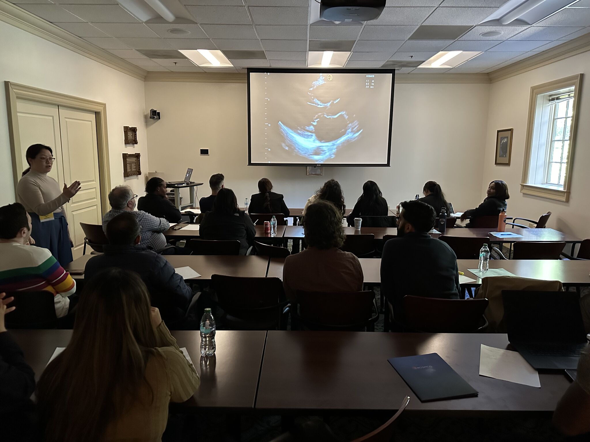
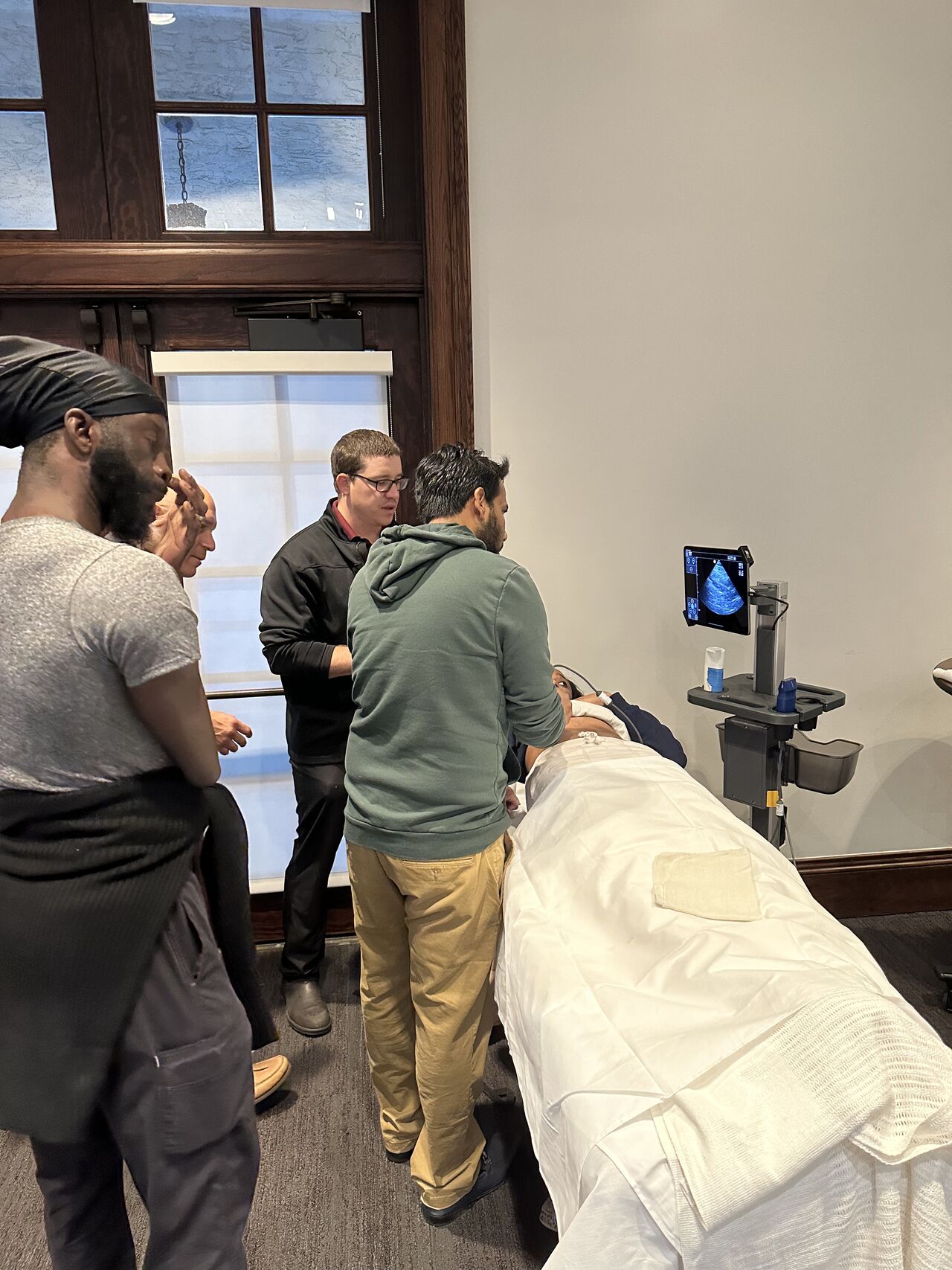
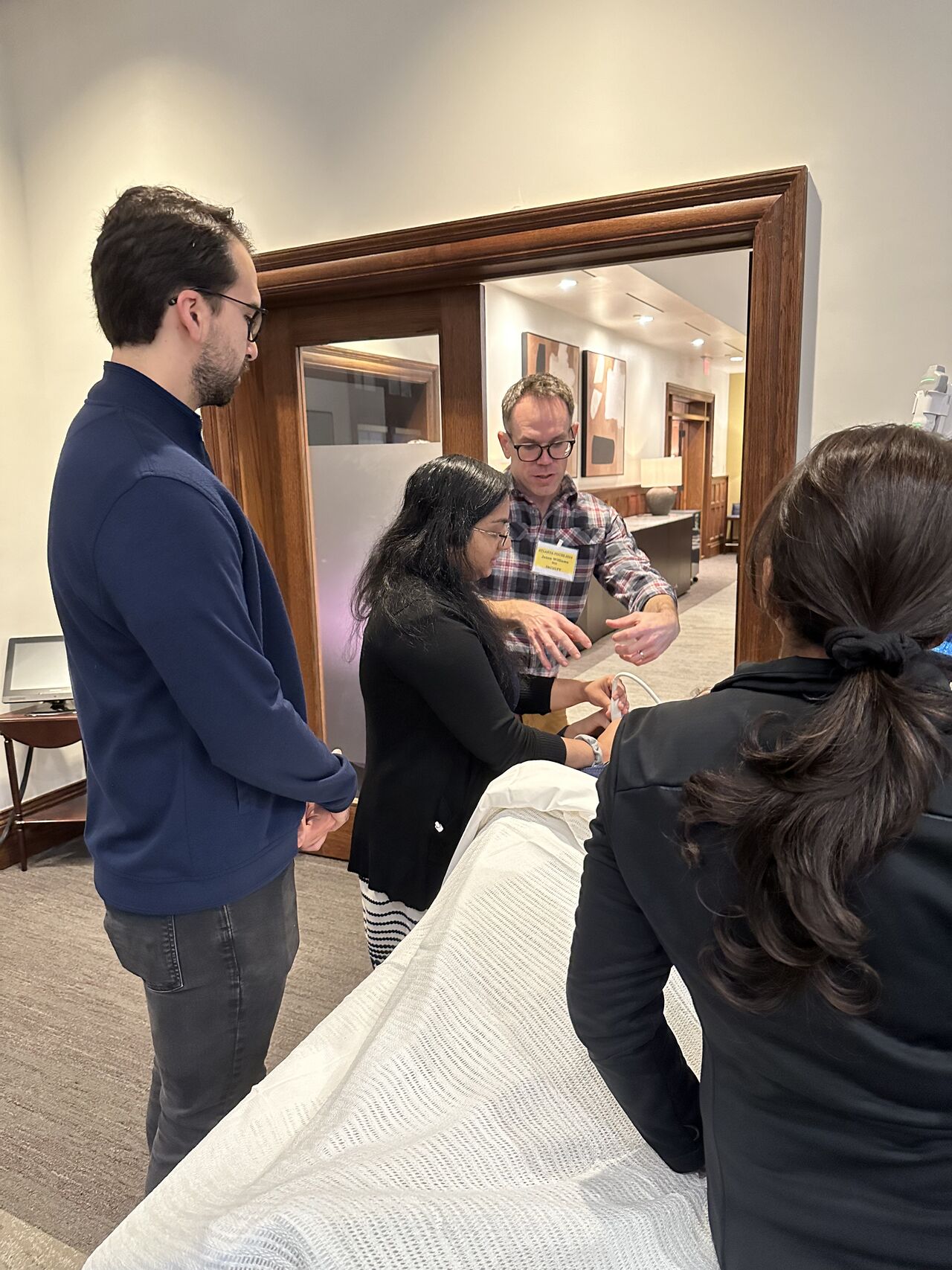
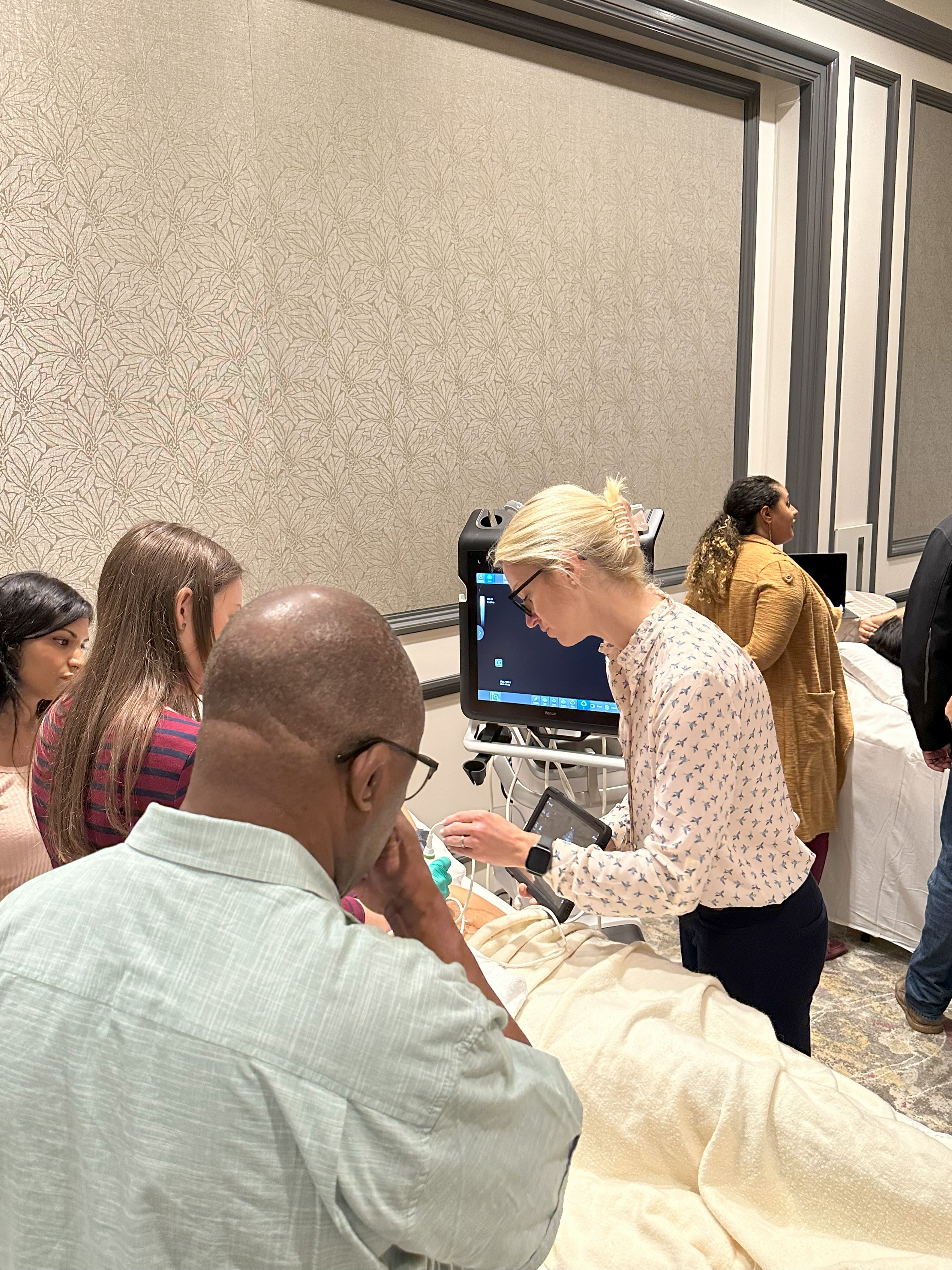
This conference is supported by Emory's Division of Hospital Medicine and is one of only seven courses approved by the Society of Hospital Medicine and American College of Chest Physicians, nationally as a course for their certificate of completion program for POCUS. The course provides attendees with hands-on training in a 2:1 learner-to-faculty ratio, didactics on POCUS for various organ systems, and clinical integration of POCUS into practice.
2025 Course Details
This year's Atlanta POCUS (Point of Care Ultrasound) Conference will take place at the Miller Ward Alumni House on November 6 and 7, 2025.
The closest hotel to this event will be the Emory Conference Center Hotel, located at 1615 Clifton Road NE
Atlanta, Georgia, 30329. You can reserve a room at the button below.
Please note that registration is now full. Please email Sheila Harris and Tiffney Stringer to be placed on the waitlist.
Course Directors:
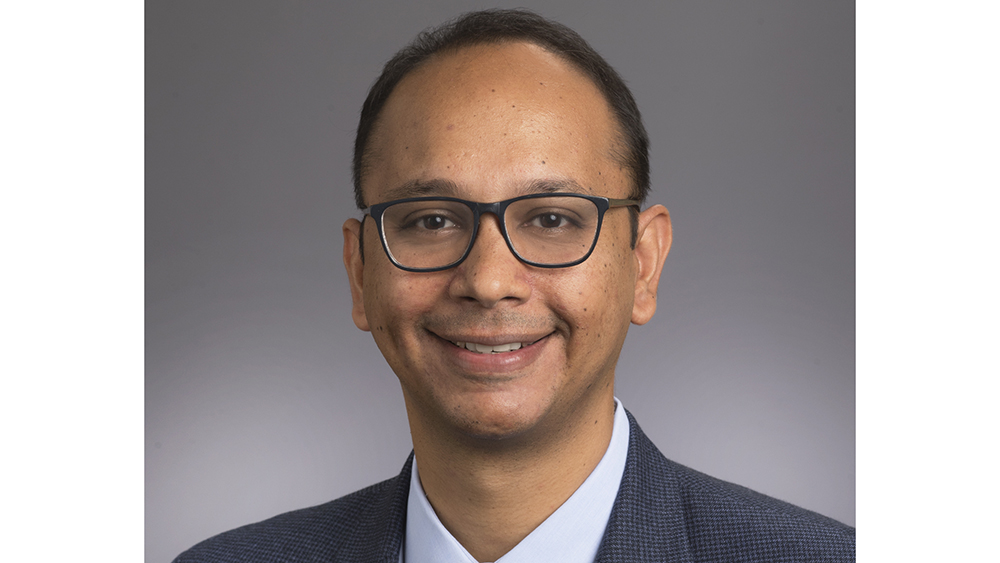
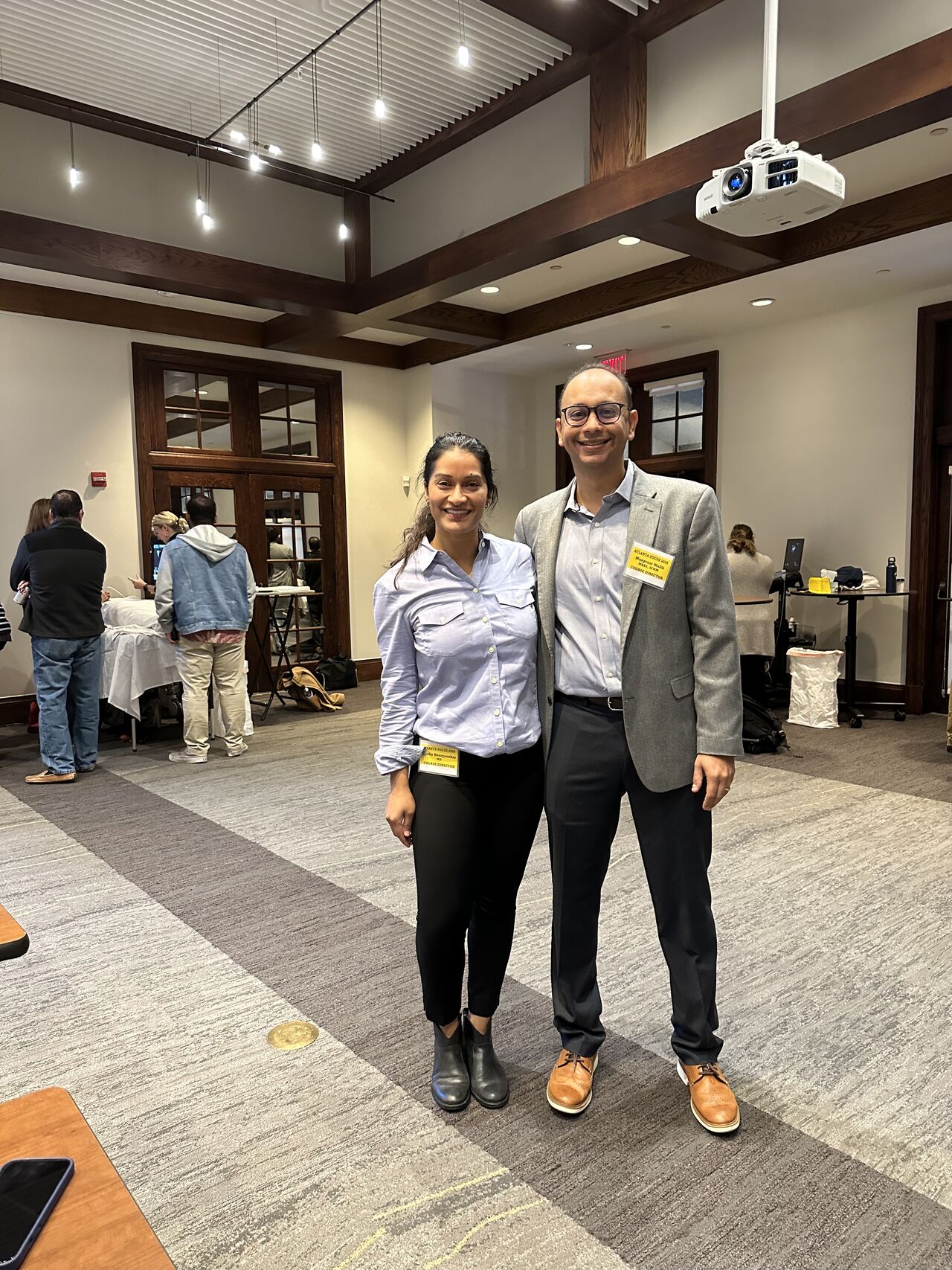
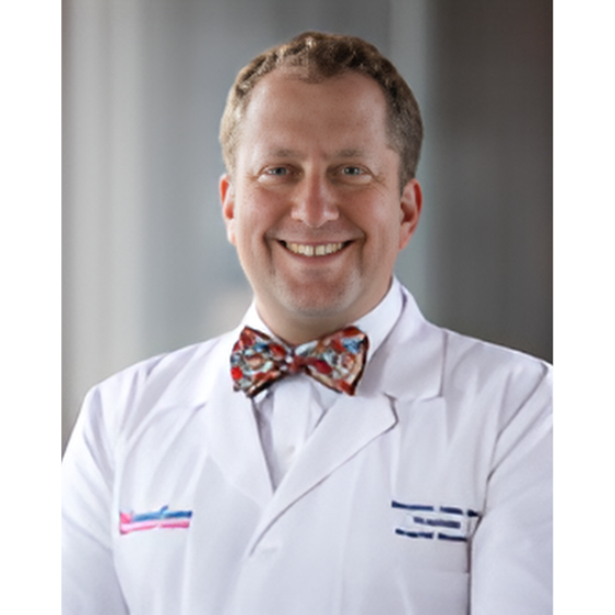
Manpreet S. Malik, MBBS, SFHM
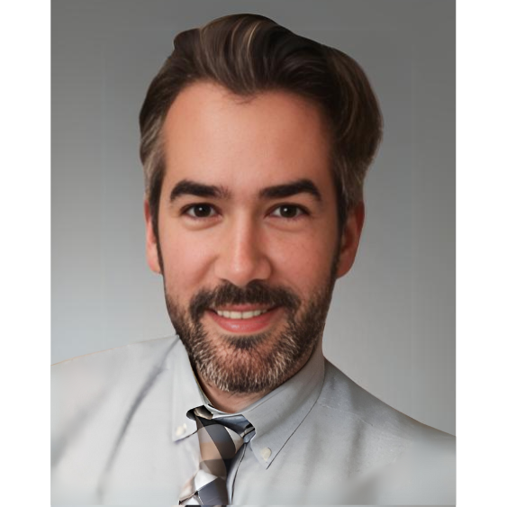
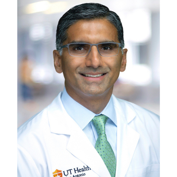
Dr. Malik is an academic hospitalist at Emory University in Atlanta. He is an Associate Professor of Medicine at Emory and clinically works at Grady Memorial Hospital. Dr. Malik is the program director for the Transitional Year program at Emory and has created and led multiple bedside ultrasound-guided procedure services, including the one at Grady. He provides faculty mentorship to residents on the POCUS Distinction pathway at Emory. Dr. Malik teaches and directs bedside ultrasound and procedure workshops for various professional societies and is the course director for Atlanta’s two-day POCUS course.
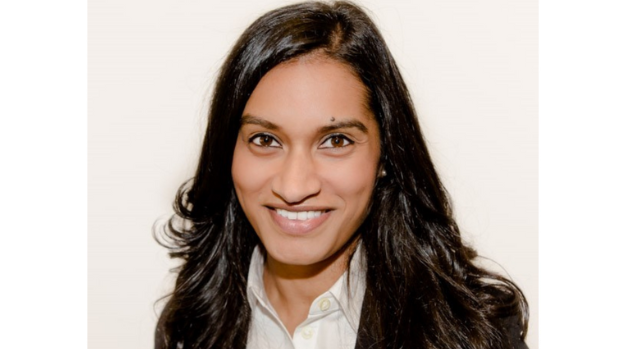
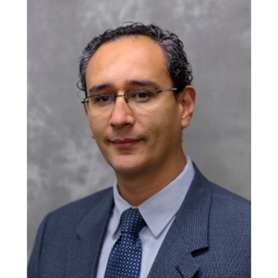
Sneha Neurgaonkar, MD
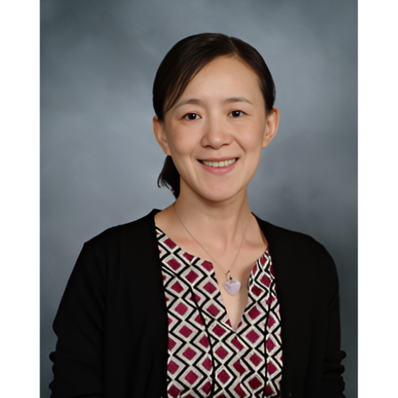
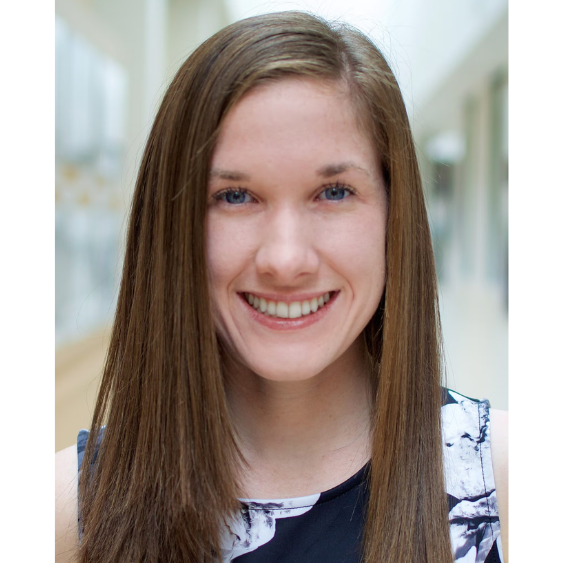
Dr. Sneha Neurgaonkar is currently an academic hospitalist and associate director of the pocus/procedure service at the Atlanta VA Medical Center. She is a faculty mentor for the clinical distinctions POCUS track for the Emory Internal Medicine residency program. Dr. Neurgaonkar is also a faculty instructor for pocus and procedure courses at regional and national conferences.
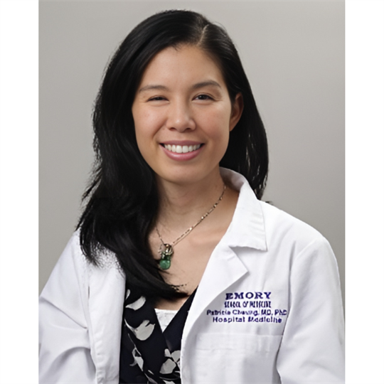
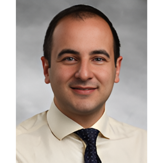
Patricia Cheung, MD
Assistant Course Director
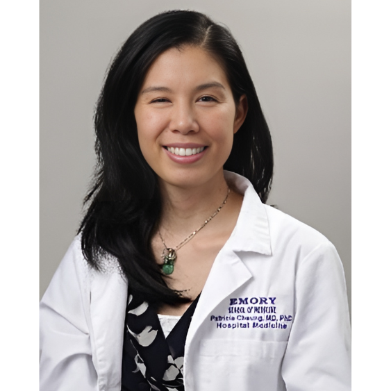
Patricia Cheung, MD PhD, is an academic hospitalist at Grady Memorial Hospital and an assistant professor at Emory University in Atlanta. She created and currently leads the POCUS curriculum for the Senior Medicine Internal Medicine Clerkship. She is a faculty mentor for the Emory Internal Medicine residency POCUS distinction program. She has served as an adjunct faculty member in the Weill Cornell Hospital Medicine Point-of-Care Ultrasonography.
Course Agenda
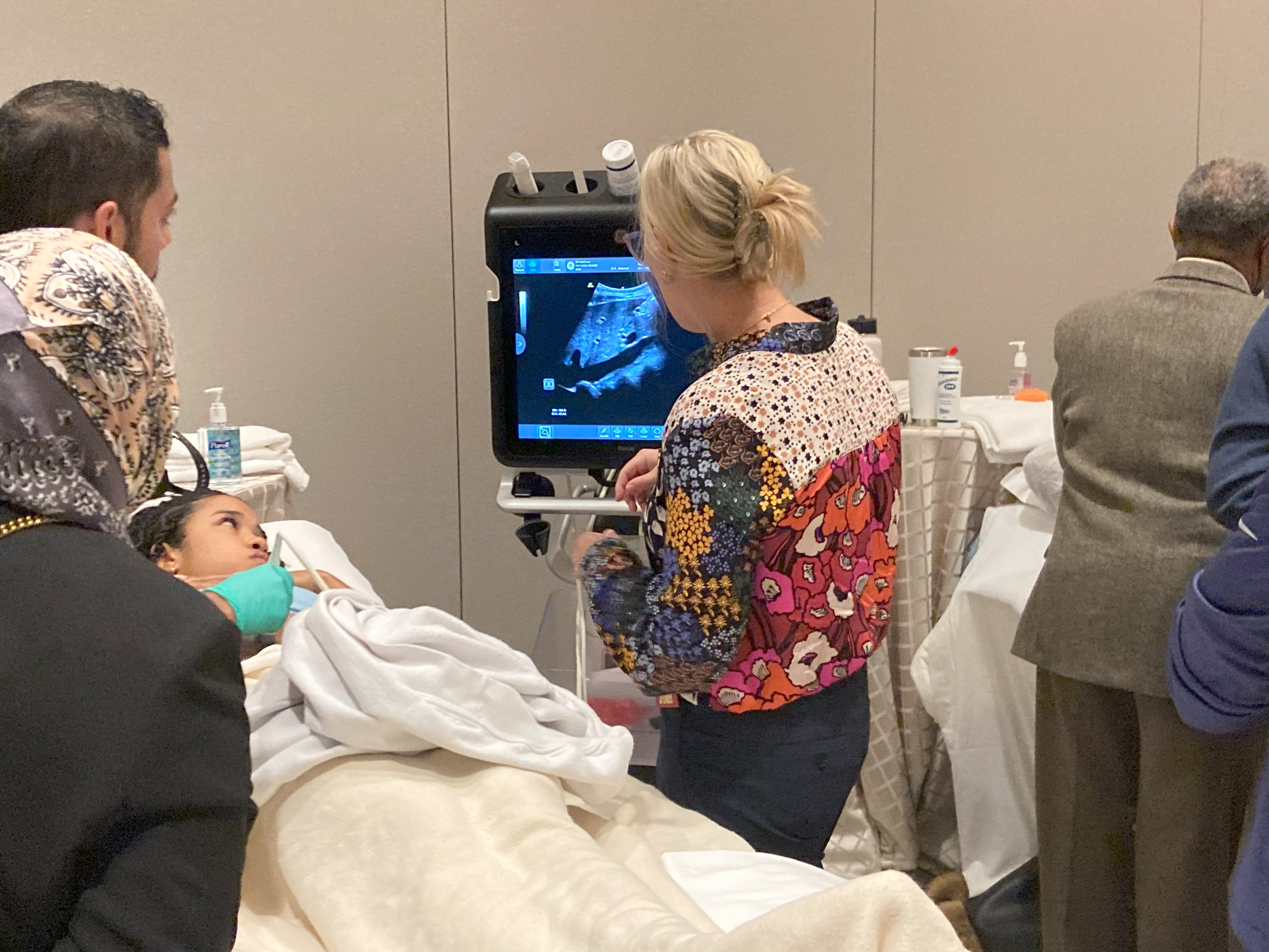
Day 1:
LUNGS, CARDIAC, DVT:
7–7:45 a.m. | Registration and Breakfast
7:45–8 a.m. | Welcome and Course Overview
8–8:15 a.m. | Ultrasound Basics
8:20–8:50 a.m. | Lung and Thoracentesis Review
8:50–9 a.m. | Break into Group A and Group B
9–9:30 a.m.| Lung scanning Group A (Scanning Room). Image Interpretation Group B (Lecture Room)
9:30–10 a.m. | Lung Scanning Group B (Scanning Room). Image Interpretation Group A (Lecture Room)
10–10:20 a.m. | DVT Review
10:25–10:55 a.m. | DVT Scanning Group A (Scanning Room). Image interpretation Group B (Lecture Room)
10:55–11:25 a.m. | DVT Scanning Group B (Scanning Room). Image interpretation Group A (Lecture Room)
11:25 a.m.–noon | Cardiac Introduction
noon–1 p.m. | Lunch
1–1:50 p.m. | Cardiac Scanning Group B (Scanning Room), Image Interpretation Group A (Lecture Room). Cardiac scanning: Attendees will Scan PLAX and PSAX at the first station and rotate at 25 minutes to Station 2 or Apical, subcostal, and IVC views.
2–2:50 p.m. | Cardiac Scanning Group A (Scanning Room), Image Interpretation Group B (Lecture Room)
2:50-3 p.m. | Break
3–3:30 p.m. | Cardiac Image Optimization
3:30–4:30 p.m. | Free scanning: Cardiac, Lung and DVT
4:30–5 p.m. | Gall bladder, Abdominal Aorta Review, and Image Interpretation
5 p.m.| Adjourn
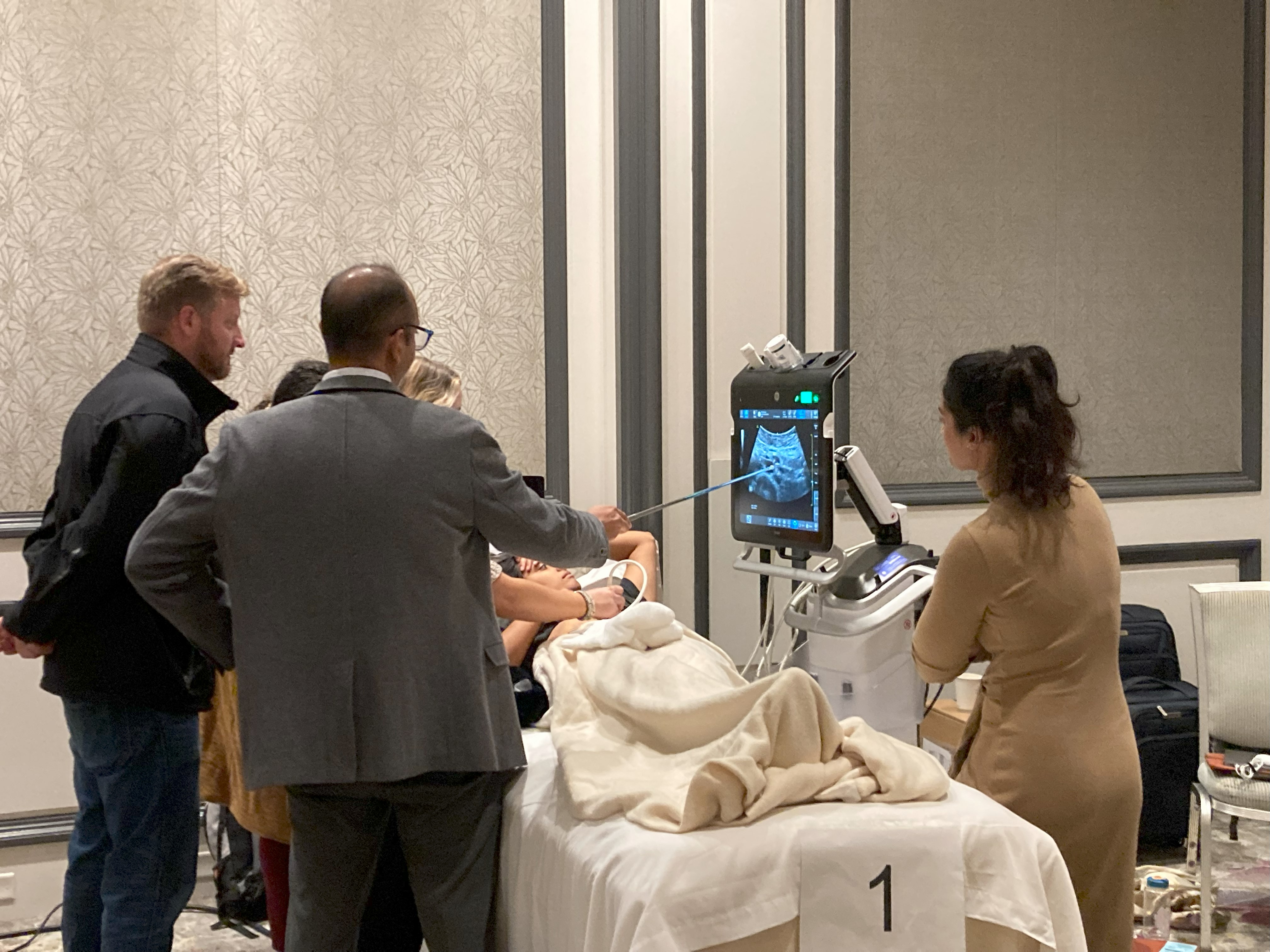
Day 2:
ABDOMINAL, FREE SCAN, VOLUME STATUS, AND CARDIAC:
7:30–8 a.m. | Continental Breakfast
8–8:45 a.m. | Gallbladder and Aorta Scanning (all learners)
8:45–9:20 a.m. | Abdomen and Paracentesis Lecture
9:20–9:50 a.m. | Abdominal Scanning (Group A)/Image Interpretation (Group B)
9:50–10:20 a.m. | Abdominal Scanning (Group B)/Image Interpretation (Group A)
10:20–10:45 a.m. | MSK, Skin, and Soft Tissue Review
10:45–11:15 a.m. | Procedural Ultrasound Review (Central line, Peripheral IV, LP, and Arthrocentesis)
11:15 a.m.–noon. | MSK and Procedural Scanning (All groups)
noon–1 p.m.| Lunch and POCUS Program Discussion with Faculty
Faculty to join attendees in the lecture room for a session on integration, devices, and curricula (45 minutes)
1–1:40 p.m. | Integration of POCUS into physical exam, evaluation of volume status, and shock
1:40–2:30 p.m. | Case-based scanning (Group A)/Image Interpretation (Group B)
2:30–3:20 p.m. | Cardiac Introduction Case-based scanning (Group B)/Image Interpretation (Group A)
3:20–3:30 p.m. | Break
3:30–4 p.m. | Post-course test and course evaluation. Wrap up.
4–4:30 p.m. | Free scan
4:30 p.m. | Adjourn