This week's Image comes from Dr. Mikaela Chilstrom. She used the linear probe to localize a soft tissue FB (Image 1) in a pt known for "subcutaneous self-insertion" of FBs.
Image 1
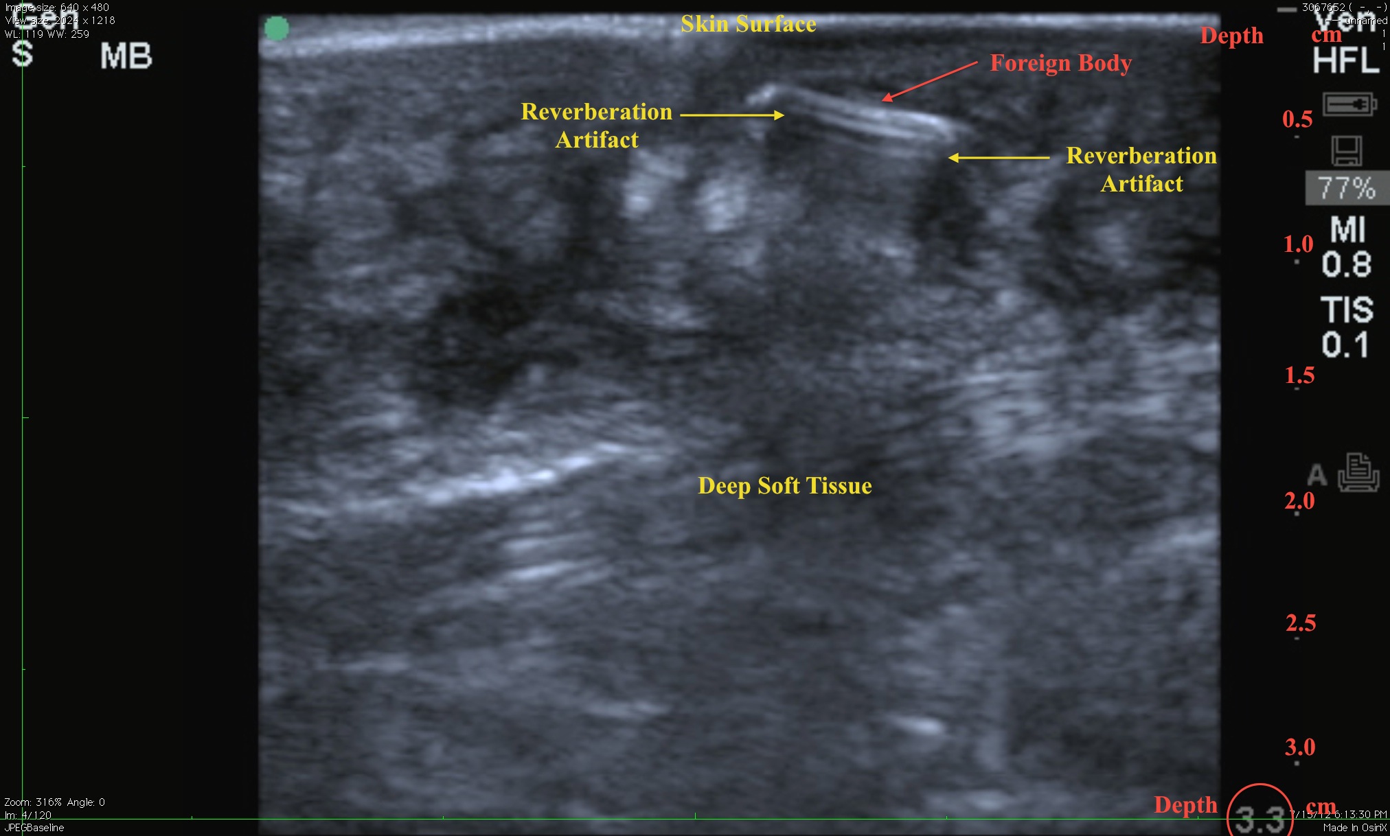
Note the hyperechoic structure with reverberation artifact beneath it - this is classic for a metallic FB. Soft tissue is best evaluated using the high-frequency linear probe. Scan in two planes perpendicular to each other to best visualize the FB as well as the surrounding tissues.
When scanning soft tissues:
- Gel is your friend. Feel free to pile it up to improve your image. If an associated wound/cellulitis/abscess exists where you want to place the probe, protect the probe and the pt by placing a Tegaderm over the probe footprint and use sterile gel (KY packet works well).
- Adjust the depth. In the image note the overall depth of 3.3 cm with dots and hashes at each 0.5 cm. The FB is located in the first 1 cm. Setting the depth slightly deeper permits visualization of structures that could make extraction difficult - if not contraindicated - in the ED.
- Stabilize the transducer. Rest the base of your hand (or ring, little fingers) on the pt to permit better control of the transducer and facilitate fine controlled movements.
Date: July 2012