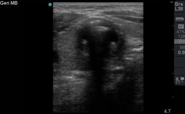
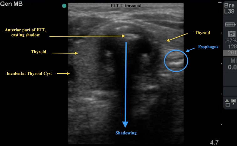
This week's Image of the Week was performed by Dr. Doug Chesson, PGY-1. He successfully captured ultrasound images confirming endotracheal tube (ETT) placement.
Ultrasound, by virtue of looking simultaneously through the neck, offers yet another method to assess ETT placement.
Performing this US in real-time begins with the trans-tracheal placement of the linear probe at the level of the crico-thyroid membrane. The probe indicator should be to the R and the tail of the probe should be angled slightly toward the head of the patient. The curvilinear probe may also be used in the same orientation. In this position, you will see a cross-section of the trachea, thyroid gland, and esophagus. The trachea has hyperechoic, non-collapsing walls with an anechoic interior. The much smaller and collapsed esophagus generally lies on the R of the screen. The trachea at this level is surrounded by the thyroid gland on both R and L sides.
Image 1

Image 2

While the patient is being intubated, a second provider can simultaneously perform the trans-tracheal US and capture the ETT as it enters the field of view. Reverberation artifact is generally noted first – when the stylet is in place. Second, once the stylet is removed, classic shadowing off of the anterior surface of the ETT is noted. In Image 2, note the hyperechoic curved density representing the ETT with distal shadowing. Alternatively, one may see the ETT enter the esophagus with the respective shadowing and reverberation artifact noted outside of the trachea.
Date: January 2012

