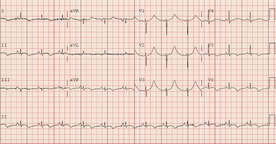
The IOW this week comes from Drs. Terry Singhapricha, Brad Wallace, and Justin Schrager who used bedside ultrasound to evaluate a patient who presented with 24 hours of constant chest pain and the EKG below.
Image 1
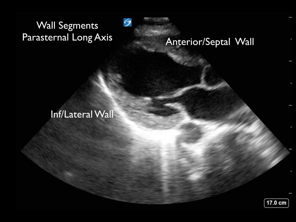
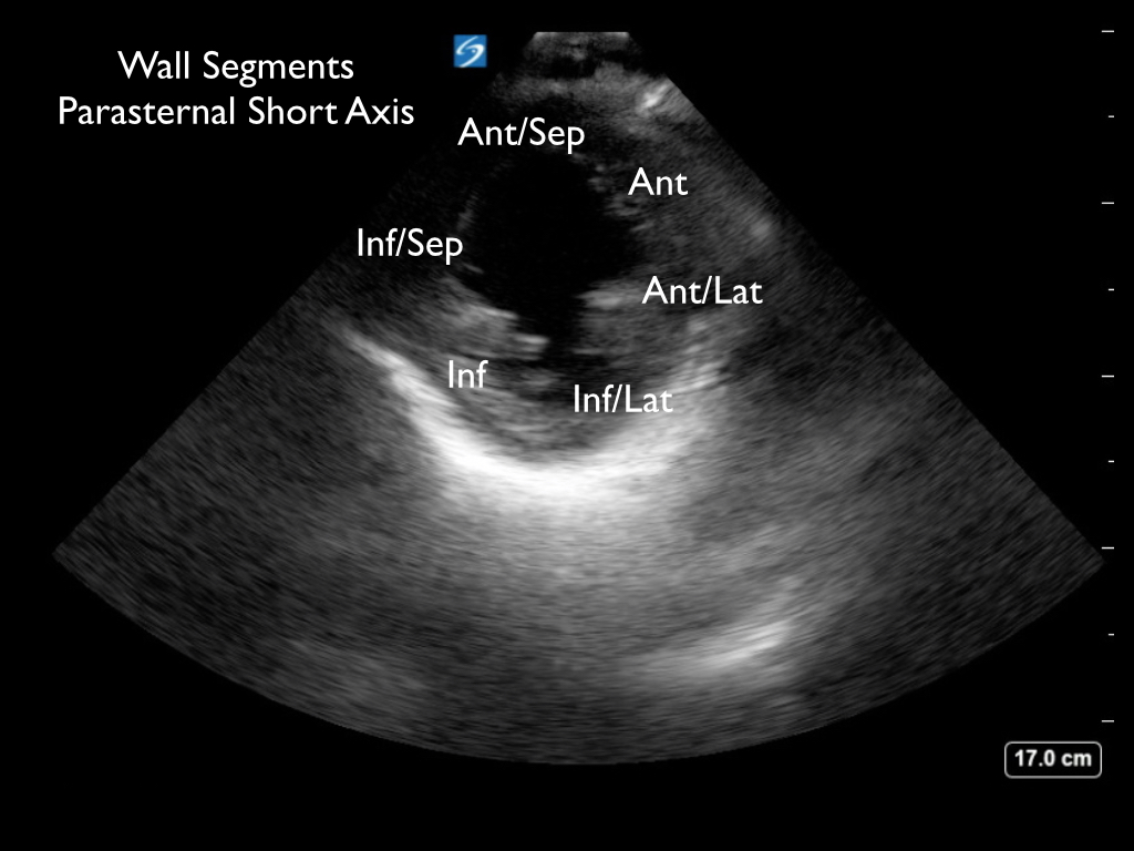
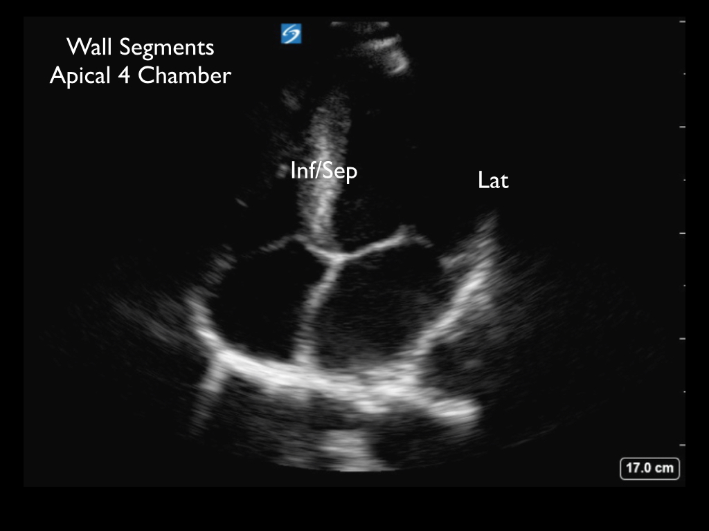
Bedside ultrasound can be useful to evaluate for wall motion abnormalities when there is concern for acute myocardial ischemia. Wall segments are labeled on the still images below.
Images
The video from their study is here.
See if you can identify the hypokinetic wall segments. Normal myocardium will thicken and move centrally into the chamber. In contrast areas of the heart with ischemia may show decreased motion, no motion, or paradoxical motion. It may be helpful to place a mouse cursor in the center of the chamber and then focus your eye on each wall segment individually to evaluate how the wall moves relative to your cursor. This patient went to the cardiac cath lab and was found to have a 100% Left circumflex occlusion. He was stented and did well.
Thanks for all of your great images this week!
Happy Scanning!
===============================
Sierra Beck MD
Assistant Professor
Department of Emergency Medicine
Emory University SOM
The IOW this week comes from Drs. Terry Singhapricha, Brad Wallace, and Justin Schrager who used bedside ultrasound to evaluate a patient who presented with 24 hours of constant chest pain and the EKG below.
Image 1
Bedside ultrasound can be useful to evaluate for wall motion abnormalities when there is concern for acute myocardial ischemia. Wall segments are labeled on the still images below.
Images
See if you can identify the hypokinetic wall segments. Normal myocardium will thicken and move centrally into the chamber. In contrast areas of the heart with ischemia may show decreased motion, no motion, or paradoxical motion. It may be helpful to place a mouse cursor in the center of the chamber and then focus your eye on each wall segment individually to evaluate how the wall moves relative to your cursor. This patient went to the cardiac cath lab and was found to have a 100% Left circumflex occlusion. He was stented and did well.