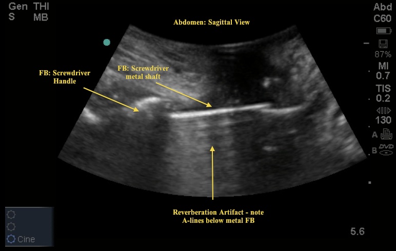
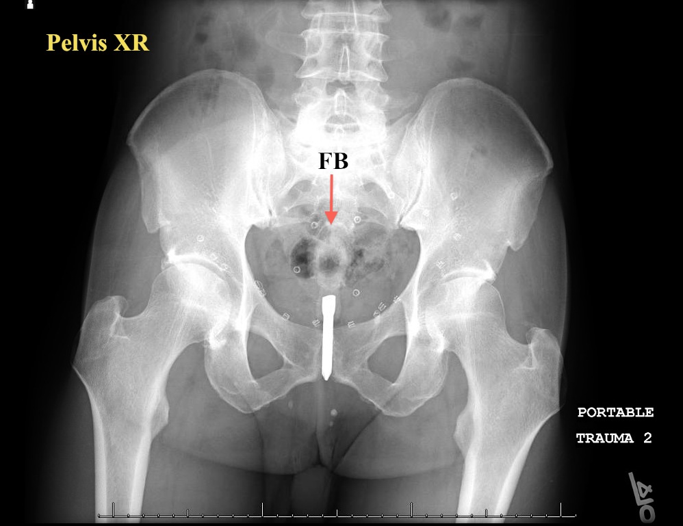
In this Image of the Week, US was used to further aid in the evaluation of a patient who presented with “constipation.” The patient was initially not forthcoming with further history. The Emory EM doctor placed the curved probe superior to the rectum, in a sagittal plane. Can you identify the cause of discomfort in this sagittal US view (Image 1)? What is the name of the US artifact seen in this image? Perhaps the XR will add some missing details (Image 2).


In brief, the patient presented with abdominal pain and the images reveal why – rectal FB. The ultrasound artifact is called reverberation and occurs when the US beams bounces off a highly reflective structure. This typically produces A-lines (horizontal lines) below the metallic structure (Image 1).
Procedural sedation and ED retrieval were successful without complication.

