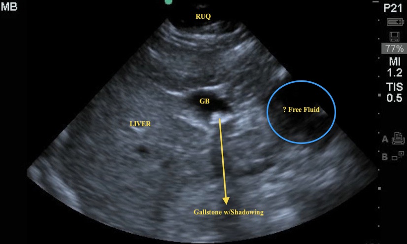
On this image, a RUQ view capturing the gallbladder, note the shadowing from a gallbladder full of stones. Is this the primary cause of pain? Note the secondary finding more toward the midline, what is that hypoechoic structure?

Image 2
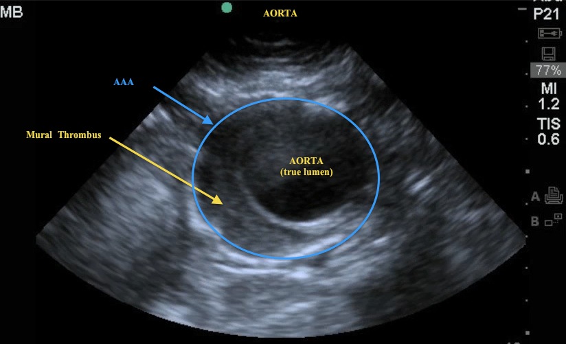
In this image, the probe has been moved to the midline from the RUQ. Note the cross-section view of the aorta. Do you see the mural thrombus? This is a classic US image for a mural thrombus in the setting of a AAA.

Image 3
On this image, note the saccular aneurysm in addition to the mural thrombus - this is a more rare type of aneurysm compared to the more common fusiform aneurysm.

Learning Points
As a reminder, to correctly image the aorta, 5 images should be obtained:
1. Proximal Aorta – cross-section with measurement of the diameter
2. Mid Aorta – cross-section with measurement of the diameter
3. Distal Aorta – cross section showing bifurcation
4. Iliacs – cross section showing individual diameter measurements
5. Longitudinal view – capturing celiac trunk and SMA.
The aorta is imaged in cross-section with the curvilinear probe, an indicator to the right, and the diameter is measured from outer wall to outer wall. The lone longitudinal image rotates the probe 90 degrees with the indicator to the head of the patient.
Author: Adam Pomerleau, MD
Date: March 2012

