Department of Biochemistry

Collaborative Projects

The Liang Laboratory seeks to exploit our structural biology (cryo-EM & x-ray crystallography) and biochemistry expertise to elucidate the molecular machinery and associated mechanisms of traditionally challenging and exciting biological systems through collaboration. We are particularly interested in interdisciplinary research in molecular cell biology, microbiology, neurobiology, and cancer biology. Please email Dr. Liang (bo.liang@emory.edu) directly for any potential collaboration suggestions. We look forward to working with you. Thank you!

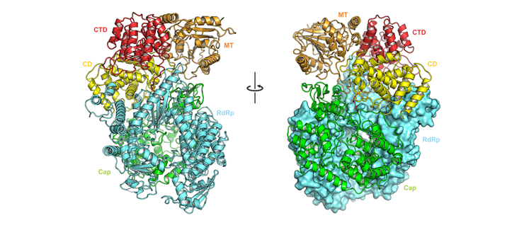
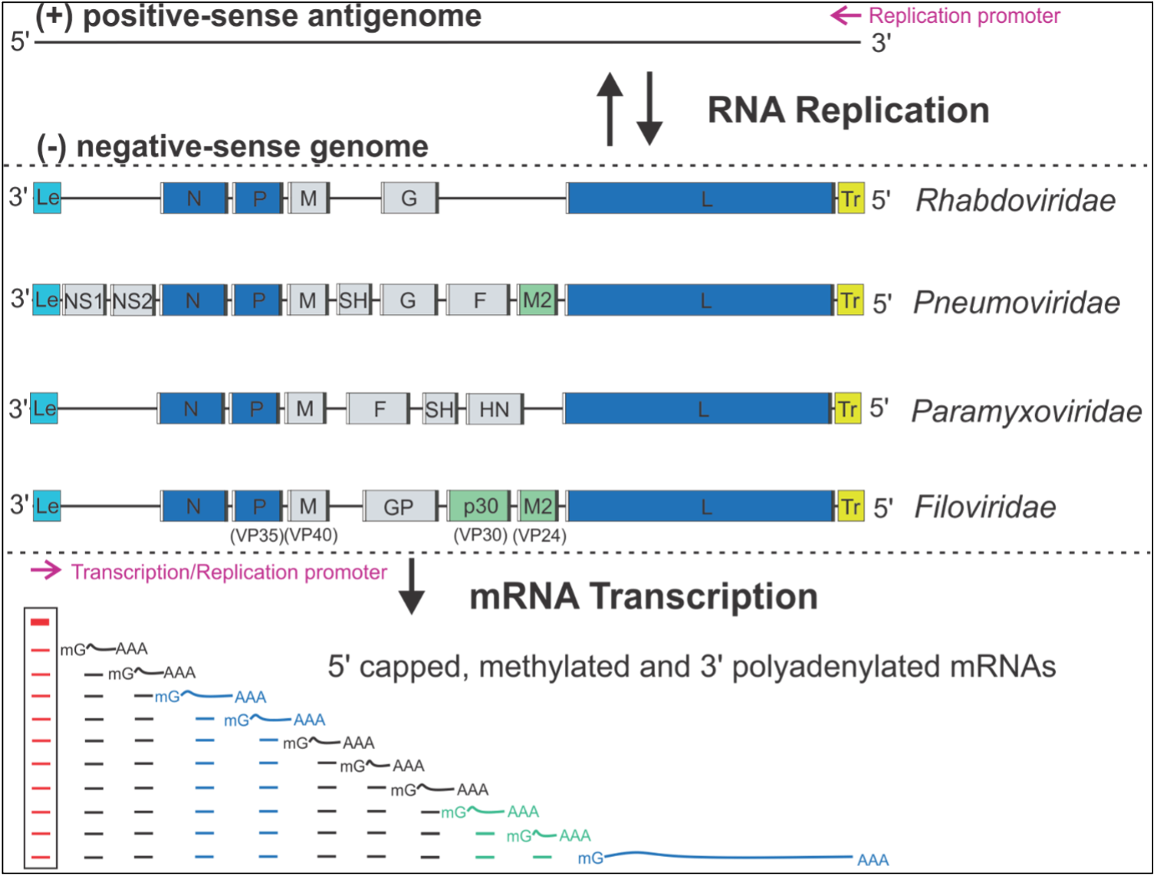
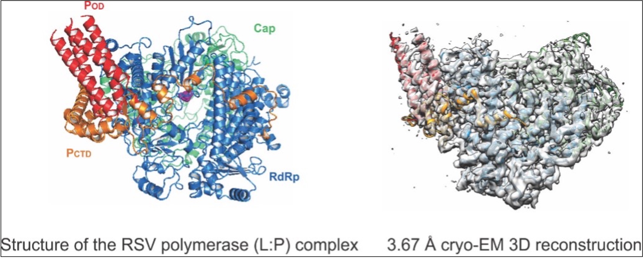
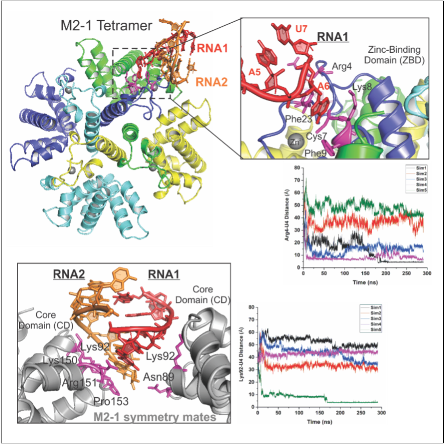
NNS RNA viruses share a common strategy of viral gene expression. NNS RNA synthesis is believed to follow the “start-stop model” of sequential and polar transcription (Fig. 1). We use RSV as a model system to delineate NNS RNA synthesis. The template for RNA synthesis is not RNA alone but rather a complex of the viral genomic RNA completely encapsidated by the viral nucleoprotein (N). This N:RNA template is copied by the viral RNA-dependent RNA polymerase (RdRP), which comprises 250 kDa large protein (L) and 27 kDa cofactor phosphoprotein (P). An additional 22kDa viral protein M2-1 is essential for full processivity in RSV. The L protein has all the enzymatic activities necessary for the transcription of the viral mRNAs, including RNA polymerization, 5’ cap addition, cap methylation, 3’ polyadenylation, and viral genome replication. Thus, L is the catalytic core of a multi-component and multi-functional RNA synthesis machine. We have successfully established protocols to prepare all key components of the RSV RNA synthesis machine. Our work on the structural and biochemical mechanisms of the RNA synthesis machinery has yielded a solid foundation for in-depth mechanistic studies. 

Liang Lab Research Image 1
Figure 1: The genome organization and RNA synthesis of Mononegavirales. The negative-sense NNS genome is depicted from the 3' end to the 5' end, showing the 3' leader (Le, cyan box), genes (gray, blue or green box) flanking with gene-start (GS, white box), and gene-end (GE, black box), and 5' trailer (Tr, yellow box). The essential genes (N, P, L) and necessary cofactors (M2 or p30) for RNA synthesis are colored in blue and green, respectively. The RNA-dependent RNA polymerase (RdRP) sequentially produces a gradient level of Le RNA (red line) and viral mRNAs (black, blue, or green line), with the attenuation of the downstream mRNAs at each gene junction. The Le RNA (red lines inside the box) remains uncapped and non-polyadenylated, while the viral mRNAs are 5' capped, methylated, and 3' polyadenylated. The lines under the Le RNA and representative viral mRNAs indicate the abundance and gradient levels of the RNA transcripts. The promoters for transcription and replication are shown with magenta arrows. (Reviewed in Liang, JVI 2020).

1. Illustration of the structure and regulation of the RSV polymerase

As the sole enzyme executing transcription and replication of RSV, the L protein is a logical target for novel antiviral drugs. However, understanding this enzyme and its cofactors at the molecular level is far from complete, impeding drug discovery efforts. The major gaps in our knowledge of the RSV RNA synthesis are the mechanistic lacking of 1) how multi-enzymatic activities of L are coordinated by its cofactor P, and 2) how L is regulated by processivity factor M2-1 during transcription to ensure that the mRNA is properly capped and polyadenylated. Importantly, robust biochemical and genetic tools are in place to dissect the enzymatic activities of L. Thus, RSV offers a clinically relevant and tractable system in which to address these knowledge gaps.  We will fill these gaps by delineating the molecular biochemistry and structural enzymology of RSV L protein with its cofactor P and processivity factor M2-1. We use cryo-EM and x-ray crystallography to obtain high-resolution images at multiple vital RSV RNA synthesis stages. Furthermore, results are expected to offer new mechanistic insight into the regulation of RSV transcription by M2-1, a processivity factor that shares structural homology with Ebola VP30. These studies will be significant within the NNS field because RSV RNA synthesis shares a similar viral gene expression strategy with NNS RNA viruses, including rabies and Ebola.


Liang Lab Research Image 2
Liang Lab Research Image 3
Liang Lab Research Image 4

2. Establish in vitro RSV RNA synthesis nucleocapsid platform 

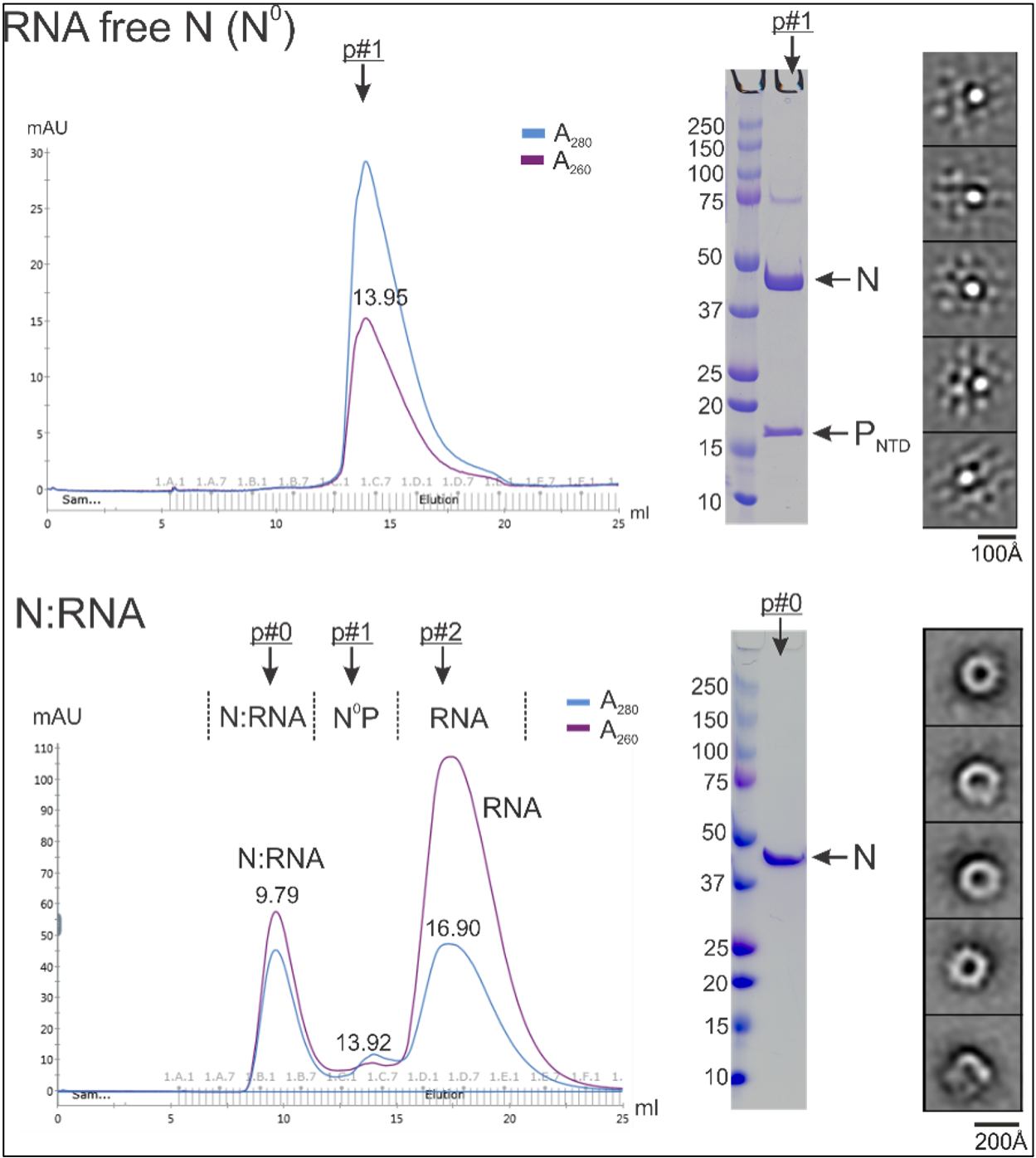
Despite the evident importance of RNA synthesis by NNS RNA viruses, an in-depth mechanistic understanding is lacking. The lack of a tractable in vitro nucleocapsid has hindered the field as a cognate RNA template for the polymerase activities. As highlighted below, the polymerase template is not naked RNA but a helical nucleocapsid (NC, N:RNA), formed by viral RNA tightly encapsidated by the nucleoprotein (N). Each RSV N coats 7 nucleotides (nts). As a result, the entire RSV genome (A2 strain, 15,222 nts) requires more than 2,100 copies of N for coating. We hypothesize that the RNA promoter sequence and the 3' end of NC's tertiary structure are vital for RNA synthesis. The polymerase must first recognize the 3' NC helical structure, displace the first few N molecules from NC and interact with the RNA promoter at the proximal 3’ terminus of the genome to initiate RNA synthesis. The successful reconstitution of the NC with essential RNA cis-acting elements is critical for the polymerase to engage the RNA template for RNA synthesis. While polymerase itself has been reconstituted in vitro, its biologically relevant N:RNA template poses a major technical hurdle, mainly because the N protein binds non-specifically to cellular RNAs to form nucleocapsid-like particles (NCLP). The primary objective of this project is to overcome this critical barrier and provide novel mechanistic insights into the NNS RNA synthesis machinery. We expect to fill this gap by designing and reconstituting an RSV mini-nucleocapsid (mini-NC) in vitro system.