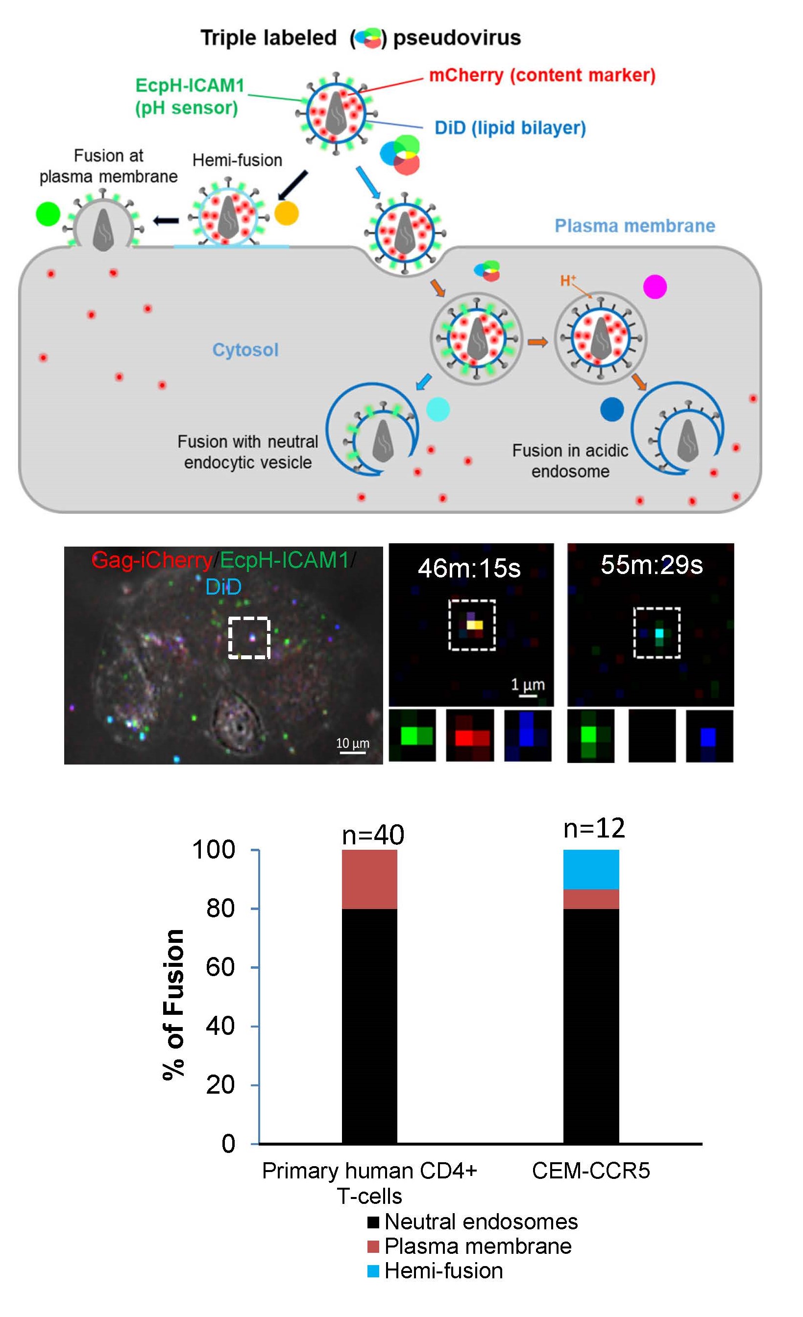
Our single virus tracking experiments revealed that HIV-1 preferentially fuses with pH-neutral intracellular vesicles but not with the cell plasma membrane of primary CD4+ T-cells. This was achieved through co-labeling of HIV-1 pseudoviruses with three fluorescent tags: viral content marker released upon fusion, virus surface-exposed pH sensor, and a lipophilic dye (Figure 1). The extent of DiD dilution upon fusion discriminates between HIV-1 fusion with the plasma membrane vs endosomes. The extent of lipophilic dye dilution upon viral fusion with the plasma membrane and the endosomal membrane allows pinpointing the sites of HIV-1 hemifusion/fusion (Movie 1). We are interested in delineating the mechanism that favors HIV-1 fusion with early endocytic compartments.


