FIBI (Fluorescence Imitating Brightfield Imaging Microscopy)
Non-Destructive Slide-Free Histology
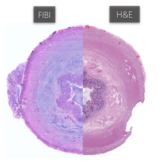
FIBI (Fluorescence Imitating Brightfield Imaging) is a slide-free histology approach that captures images directly from tissue surfaces using fluorescence-based methods, eliminating the need for traditional tissue preparation steps like fixation, paraffinization, and sectioning. This method is fast, cost-effective, and shows concordance with traditional H&E-stained sections. FIBI is particularly useful for rapid histological diagnostics and intraoperative guidance, preserving tissue for further downstream tissue analysis.
Figure 1. Comparison of the appendix with FIBI imaging (left) and traditional FFPE H&E (right).
DUET (Dual-mode Emission and Transmission Microscopy)
Detect More with Less: Generation of Virtual Special Stains from H&E Slides
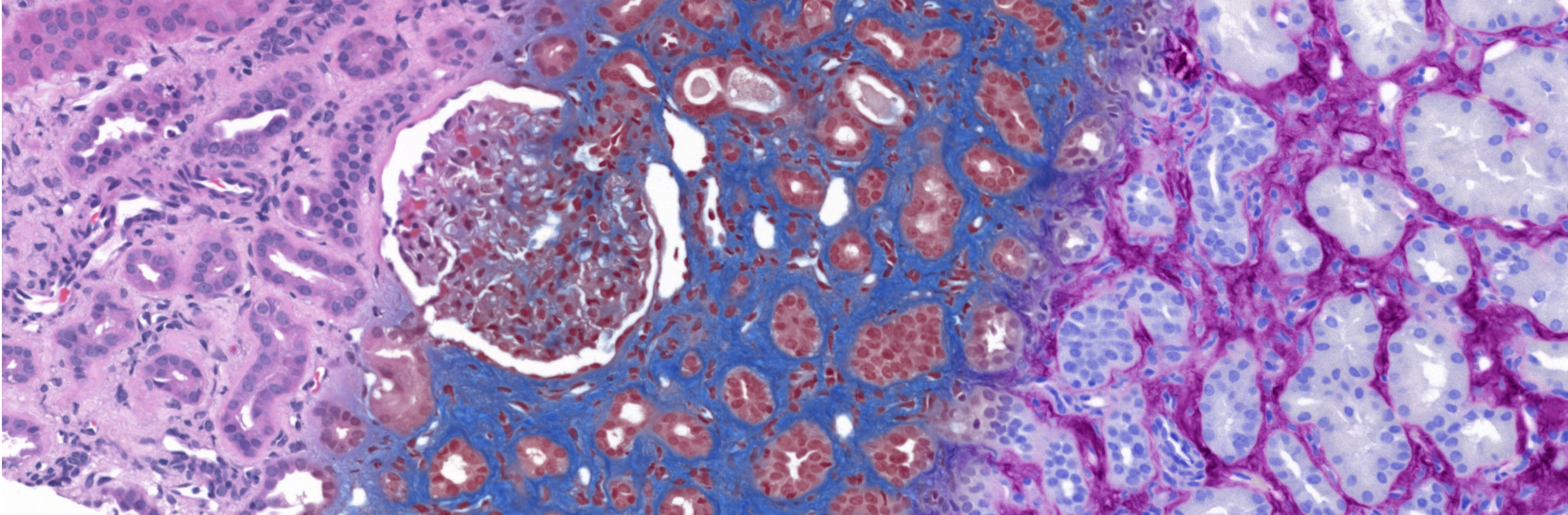
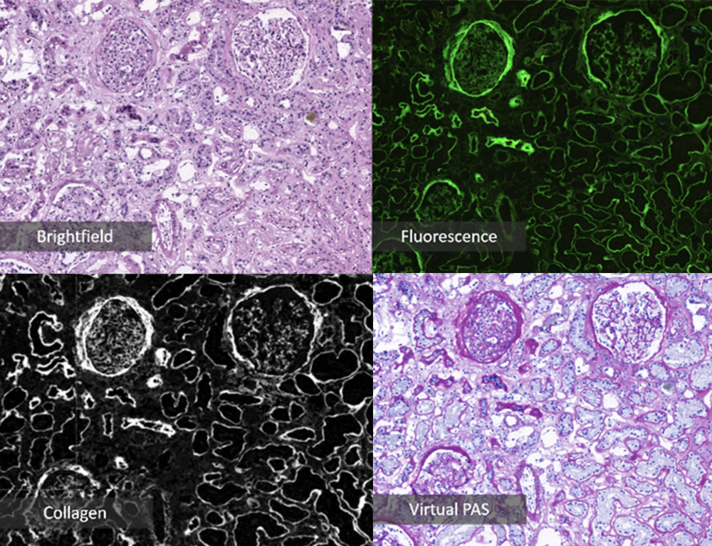
DUET is a specialized digital slide scanner that extracts more (and often ignored) information from H&E-stained slides using both brightfield and fluorescence microscopy. Fluorescence images from DUET highlight collagen and elastin features, which can be converted into virtual stains, such as Trichrome or PAS, reducing the need for additional histological staining. Virtual stains on frozen H&E slides address the time constraints of traditional special stains and are well-suited for translation to provide intraoperative guidance to pathologists. DUET-generated virtual Trichrome and PAS images are being evaluated as a rapid, effective tool for assessing kidney graft viability. Additionally, leveraging fluorescence imaging and machine learning, we are exploring the roles of collagen and elastin in diseases like chronic kidney disease and cancer.
Figure 2. DUET image panel for virtual PAS generation. Brightfield and fluorescence images are used as inputs to generate a virtual collagen mask. Collagen masks are then overlayed with the original brightfield H&E image to generate a virtual PAS stain.
GigaFIBI (FIBI, but bigger and better)
Non-Destructive Slide-Free Histology for Whole Organ and Tissue Imaging
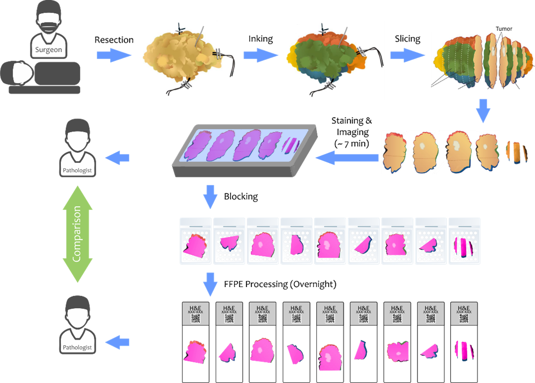
GigaFIBI, the next iteration of FIBI, is designed to provide high-resolution images of large tissue specimens (up to 100 x 100 mm²) for real-time intraoperative guidance. Breast conservation surgery (BCS) aims to remove tumors while preserving healthy tissue, making accurate margin assessment critical. Currently, lumpectomy margins are evaluated using H&E slides, which take days to process, delaying decisions and often leading to repeat surgeries. This project tests the feasibility of GigaFIBI in BCS by comparing its diagnostic accuracy to traditional H&E evaluation. If validated, GigaFIBI could enable real-time margin assessments during surgery, reducing repeat procedures and improving patient outcomes.
Figure 3. Proposed workflow for the GigaFIBI feasibility and validation study. Breast cancer tumor margins will be evaluated by pathologists and compared using GigaFIBI images to traditional FFPE H&E slides. Surgically resected tissue will be imaged with GigaFIBI before blocking, but after inking, breadloafing, and staining with H&E. After imaging, tissue is returned to routine histology and preserved for downstream testing.