School of Medicine Homepage
Emory University Shield
  • About
    • Our Vision
    • Academic Departments
    • Our Leadership
    • Health Care Partnerships
    • Diversity, Equity, & Inclusion
    • Location
    • Supporting Our People
    • Our Faculty
    • History
    • News
    • Events
  • Education
    • Degree Programs
    • Academic Departments
    • Residents & Fellows
    • Postdoctoral Training
    • Continuing Medical Education
    • Admissions
    • Cost and Financial Aid
    • Student Resources
  • Research
    • Research Innovation
    • Conducting Research
    • Core Facilities
    • Research Centers
    • Research Training
    • Academic Departments
    • Find a Researcher
    • Research News
  • Clinical Experience
    • Where We Work
    • Caring for a Diverse Population
    • Faculty Clinical Experience
    • Advanced Patient Care
    • Academic Departments
    • Community Learning
  • Giving
  • Directory
  • School of Medicine Home
  • Emergency Medicine
  • Emergency Medicine Sections
  • Ultrasound
  • Image of the Fortnight
  • Soft Tissue, Vascular, MSK
  • Playing tennis and you feel a pop?

Playing tennis and you feel a pop?

Ultrasound
  • Ultrasound Education
  • Faculty
  • Cases of the Month
    • Abdomen
    • Cardiac
    • IVC
    • Miscellaneous Highlights
    • Procedure Highlights
    • Lung
    • OB/GYN
    • Trauma
    • Soft Tissue, Vascular, MSK
    • Ocular Highlights
  • Related Resources
  • Contact Us

The IOW this week comes from Dr. Nicole Franks. Her patient is a 48-year-old male who presented to the ER with ankle pain and difficulty with ambulation after an injury while playing tennis. He reports he heard a pop when the injury occurred.

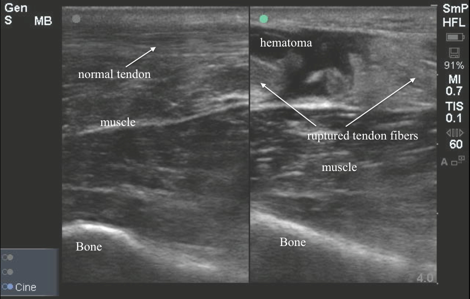
Bedside ultrasound below was obtained. To get this image place the patient in a prone position. The high-frequency linear probe is placed over the Achilles tendon in a long axis orientation. Normal tendons have a fibular appearance with many tightly packed hyperechoic linear echoes as seen on the left. In the short axis, if you rotate the probe 90 degrees, these echoes will take on a stippled appearance. In the image to the right, we see a large hypoechoic gap between tendon fibers which represents hematoma. The tendon fibers are irregular and contracted. If you squeeze the calf during your exam this defect will enlarge. If you are not sure what you are seeing its always helpful to look at the contralateral normal tendon. Achilles tendon rupture is typically a clinical diagnosis, but findings can be subtle and the diagnosis is missed in up to 20% of cases. Ultrasound can be a useful bedside adjunct.

In this case, the diagnosis of Achilles tendon rupture was made rapidly at the bedside, the patient was splinted, and orthopedic follow-up was arranged. 

Image 1

achillestendonrupture

Assistant Professor

Ultrasound Fellowship Director
Department of Emergency Medicine 
Emory University SOM

6/18/2014

Ultrasound
  • Ultrasound Education
  • Faculty
  • Cases of the Month
    • Abdomen
    • Cardiac
    • IVC
    • Miscellaneous Highlights
    • Procedure Highlights
    • Lung
    • OB/GYN
    • Trauma
    • Soft Tissue, Vascular, MSK
    • Ocular Highlights
  • Related Resources
  • Contact Us
Soft Tissue, Vascular, and MSK Case Collection
Emory University Homepage

Contact & Location

Emory University School of Medicine
100 Woodruff Circle
Atlanta, GA  30322 USA
  • Contact Us
  • Maps & Directions
  • Careers

Information For

  • Alumni
  • Faculty & Staff
  • Patients
  • Residents & Fellows
  • Students
Support the School of Medicine School of Medicine Intranet
facebook facebook twitter twitter linkedin linkedin instagram instagram youtube youtube
© 2018 Emory School of Medicine
  • Privacy Policy
  • Emergency Information