This week's image is brought to us by Drs. Dilani Weerasuriya and Christine Keyes, who used the ultrasound to confirm the diagnosis of Achilles tendon rupture in a patient who presented to the ED for sudden onset of ankle pain. Achilles tendon ruptures can be easily missed, as the presentation can appear similar to simple ankle sprains. Ultrasound can reliably help you to diagnose an Achilles tendon rupture, and can even distinguish a partial tear from a complete tendon rupture.
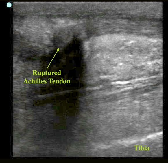
The Achilles tendon is easy to image with ultrasound. To perform this scan, it can be helpful to place the patient prone with the foot hanging over the edge of the bed. Start scanning distally at the attachment of the tendon to the calcaneus. Keep your probe oriented parallel to the leg so a long axis view of the tendon can be seen. Normally the Achilles is very superficial, less than 5mm from the skin surface, so be sure to decrease your depth if you are having trouble identifying structures. The intact tendon appears linear, fibrillar, and echogenic (Image 1). As you track proximally, the fibers of the tendon will merge into the gastrocnemius muscle. A tendon rupture will appear as a hypoechoic defect within the tendon. In Image 2, we can see a complete rupture of the tendon. Partial tendon ruptures can be more subtle appearing, as only a small dark defect in the body of the tendon. Dynamic scanning can make this defect more clear. To do this, try squeezing the calf or dorsiflexing the ankle while performing your scan, and the hypoechoic area will widen as the tendon is stretched.
Image 1
Image 2
Date: June 2012