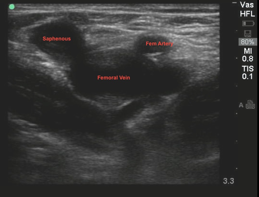
This week's image of the week comes to us from Dr. Adam Pomerleau, who used bedside ultrasound to diagnose an acute DVT. His patient was a 28yo female on oral contraceptives who presented to the ER for unilateral leg swelling and pain. To do this exam the linear probe is placed in the groin at the site where you would normally insert a central line. Your key landmark is the site where the saphenous vein empties into the femoral vein. You must capture this landmark for a complete study. Direct compression is applied to the vein with the ultrasound probe. In a normal study, the anterior and posterior walls of the vein should come together completely. You can tell that you have applied enough pressure when the artery is also slightly compressed. The image below was obtained with pressure applied and the vein remained distended. See the YouTube link for a video image.
Sometimes you will see echogenic material within the vein, but new clot may be completely anechoic, and only identified by abnormal compression. When performing the study make sure you have identified the deep veins of the groin correctly. You can be easily faked out by a superficial vein, lymph nodes, or arteries if you are not careful.
Image 1

Case Video
Date: August 2013

