December 2020
by Amanda Haan, Emergency Ultrasound Fellow
66-year-old male presented with worsening shortness of breath over the last week. Further review of systems revealed the patient had associated nausea, vomiting, and diarrhea during this time. Of note, the patient had been recently admitted to the hospital for an exacerbation of congestive heart failure. On arrival, he was found to be febrile at 38.5C, tachycardic in 140s with irregularly irregular rhythm, hypotensive with MAP in 50s, respiratory rate at 24, and hypoxic on RA to 84%.
Thoracic POCUS shown below revealed concern for subpleural consolidations, focal B lines, and irregular pleural lining.
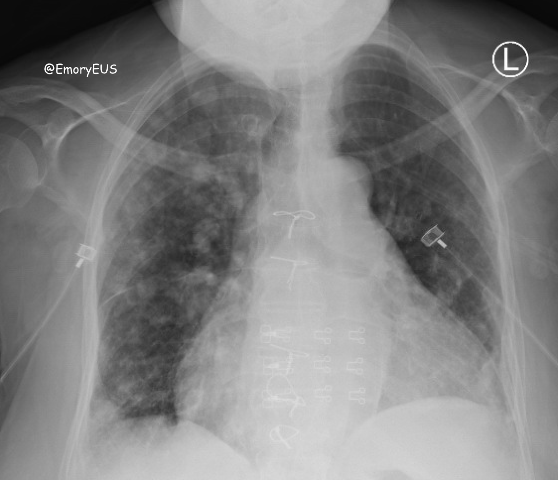
Chest xray as seen below was read as with increased nodular opacities bilaterally which draws concern for infectious process.
Upon admission, patient continued to further decompensate and required intubation. COVID testing resulted the following day and was negative. Cultures which were drawn while in ED came back positive and later a CT chest performed showed concern for septic emboli. CT chest shown below. Echo later confirmed presence of valvular vegetations.
Discussion
In the current health state of 2020, common practice across hospital systems is to assume all patients could be infected with COVID-19 to ensure that all healthcare professionals are taking the necessary precautions to protect themselves as well as subsequent patients. Thus, in patients presenting with cough, fever, or shortness of breath, COVID19 is always considered as a differential diagnosis. Among the ultrasound community, lung ultrasound in suspected COVID19 patients has been a hot topic of discussion both in diagnosis as well as in ability of providers to risk stratify patients for disposition.
Multiple protocols for lung ultrasound in COVID19 have been proposed. One study evaluated an expanded protocol of 14 anatomic areas in comparison to a limited exam of focused anterior, lateral and posterior evaluation and found that a more limited appropriate lead to missing involved lung fields (2).
It is important to note that the classic findings of COVID19 pneumonia on ultrasound are nonspecific (1). The typical findings have been described as (1, 2, 4):
-
thickening of the pleural line
-
pleural line irregularity
-
subpleural consolidations
-
B lines (focal, multifocal, confluent/watershed)
-
focal, trace pleural effusions
These findings are frequently localized to the middle and lower lung fields in the lateral and posterior distribution (4). As the disease progresses, the anticipated changes progress as well. Early COVID is typically associated with focal B lines and pleural thickening, which become more confluent with associated subpleural consolidations at later stages of the disease (4).
However, it is important to remember that in lung ultrasound these findings can be seen in multiple diagnoses and are not specific to COVID19 (1, 3), such as in this patient case of septic emboli.
Other differentials with similar findings on ultrasound include:
-
other viral pneumonias
-
lobar pneumonia
-
pulmonary edema
-
pulmonary infarct
-
pulmonary fibrosis
-
inflammatory lung disease
-
atelectasis
Even in a time of assuming all of our patients have COVID19 to ensure we maintain situational awareness for our protection as well as others, be sure to maintain a broad differential and avoid anchoring bias.
References
-
Yang Y, et al. “COVID-US: A simplified approach to cardiopulmonary ultrasound in suspected and confirmed COVID-19 patients in surge crisis.” Australasian Society for Ultrasound in Medicine. May 2020. 23 (2); 96-102.
-
Smargiassi A, et al. “Lung Ultrasound for COVID-19 Patchy Pneumonia: Extended or Limited Evaluations?” American Institute of Ultrasound in Medicine. 2020; 9999: 1-8.
-
Khalili N, et al. “Lung Ultrasound in COVID-19 Pneumonia: Prospects and Limitations.” Academic Radiology. July 2020. Vol 27, No 7. P 1044-5.
-
Buda N, et al. “Lung ultrasound in the diagnosis of COVID-19 infection – A case series and review of the literature.” Advances in Medical Sciences. 2020. 65; 378-85.