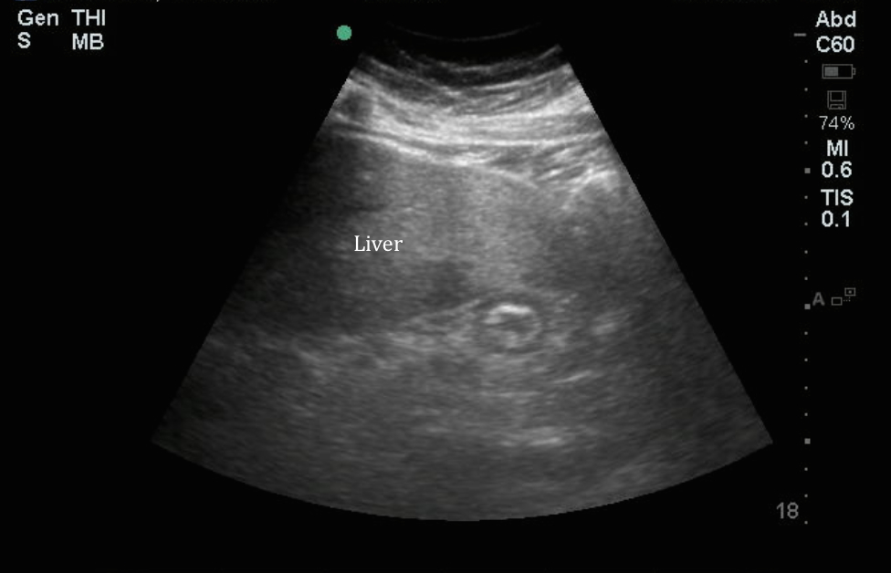
The IOW this week identified a small structure medial to the liver that is a common cause of confusion on point-of-care ultrasound. Often misidentified as the gallbladder, or a mass, the structure below can be identified by the characteristic alternating hyperechoic and hypoechoic layers.
Image 1
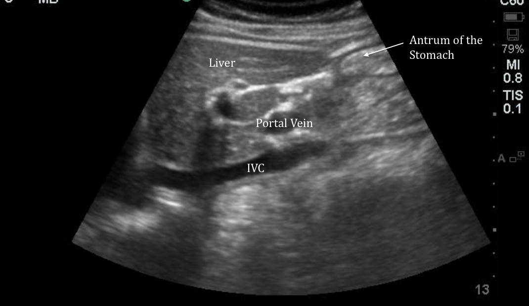
This is the antrum of the stomach. Normal bowel has a laminar appearance with alternating light and dark layers. In the clearest images 5 layers of bowel can be identified. From inside to out:
1. gastric contents and mucosa (typically bright)
2. deep mucosa/ muscularis mucosa (grey)
3. submucosa (bright)
4. muscularis propria (dark)
5. serosa (thin and bright)
If you hold the probe still and watch a section of bowel, you should be able to appreciate peristalsis in the small bowel and stomach. Normal bowel should also be compressible with pressure of the probe. Air within the bowel may cause dirty shadowing obscuring structures posterior to it. The image below shows the appearance of the antrum of the stomach in long axis. It is helpful to know what normal anatomy looks like so you don’t confuse it for pathology.
Image 2
If you want to geek out to some more bowel ultrasound this article is a nice overview… Nitin Chaubal, Manjiri Dighe, Mohit Shah, and Jyoti Chaubal. Sonography of the Gastrointestinal Tract. JUM January 2006 25:87-97
Sierra Beck MDAssistant Professor
Department of Emergency Medicine
Emory University