Image of an adult patient who presented with abdominal pain, nausea, and vomiting.
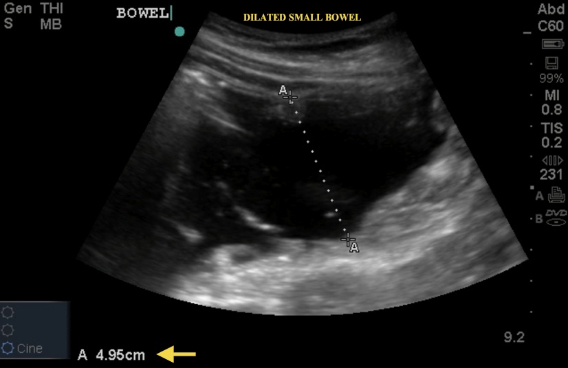
In image 1 below, notice the dilated bowel >4cm. small bowel dilated >2.5 cm is highly suggestive small bowel obstruction.
Image 1

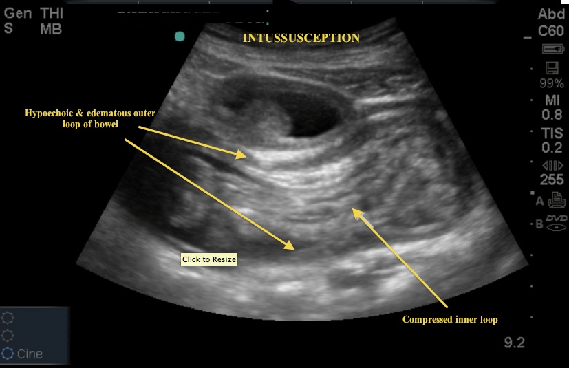
However, look at Image 2. Does this image give you an idea of what is causing the obstruction?
Image 2

This is a case of adult intussusception. Recall the textbook presentation of intussusception is abdominal pain, currant jelly stools, and a palpable "sausage-like" mass in the RUQ of a toddler-aged child. In children, intussusception is the most common cause of intestinal obstruction at age 3 mo-6 yrs. In adults, the diagnosis is rare, occurring in 1-3% of cases of bowel obstruction. The classic US sign of intussusception is the bull's eye or "target sign." Unfortunately, that cross-section view is not seen here. Rather, we see another common view - bowel telescoping inside itself (Image 2). Note the thickened hypoechoic rim representing the edematous outer bowel and the compressed inner loop. To obtain these images the curvilinear probe was used. If a mass is palpated this area should be scanned first and images obtained in two planes. If there is no mass, the entire abdomen can be scanned in a systematic fashion.
Date: September 2012

