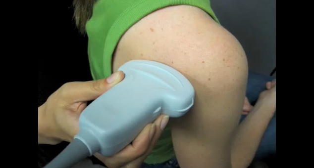
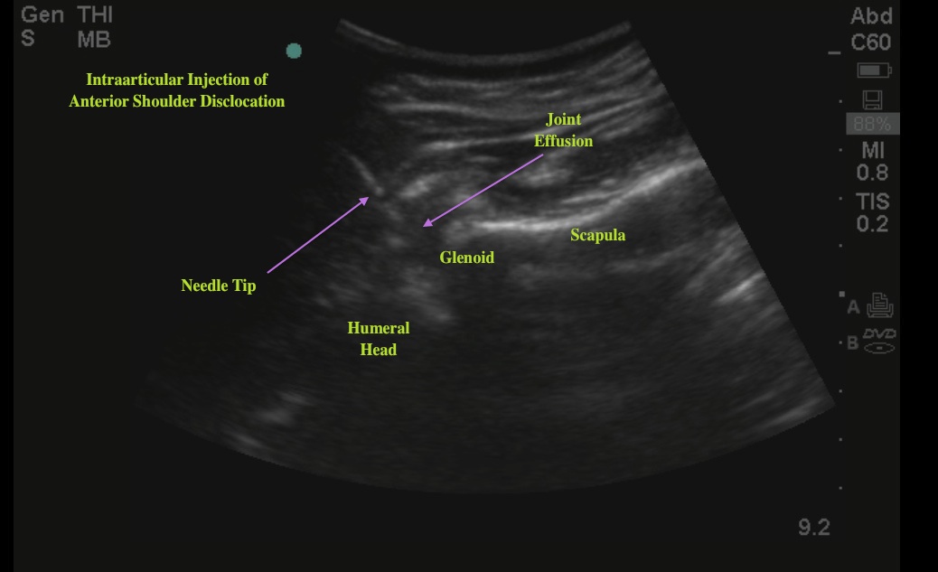
This week’s image is a real-time, ultrasound-guided, intraarticular local anesthetic injection prior to reduction of an anterior shoulder dislocation brought to us by Kyle Brown, Gail Ferek, and John Lemos. The shoulder is seen here from a posterior approach. Typically for superficial structures, we use the high-frequency linear probe, but for the shoulder, the curvilinear probe gives a wide-angle view that can help us orient to the anatomy. The probe is placed on the posterior shoulder parallel to the scapular spine with the indicator directed laterally (Image 1). The needle enters from the lateral aspect of the shoulder beneath the acromion so it can be seen in plane as it advances into the joint. A pop will be felt as your needle passes through the supraspinatus tendon.
Image 1

Image 2

This patient had great anesthesia with the intraarticular injection and was easily reduced with scapular manipulation. The reduction was confirmed with a repeat ultrasound at the bedside that showed the humeral head articulating with the glenoid with subtle internal and external rotation of the shoulder. This is a great technique that is easy to learn. It can save you the time and potential complications of procedural sedation.
Date: October 2014

