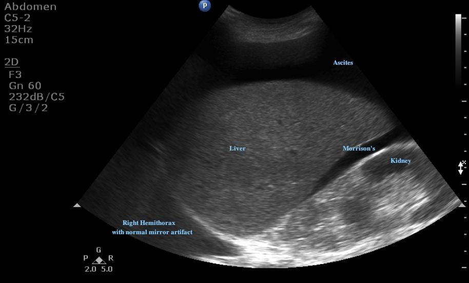
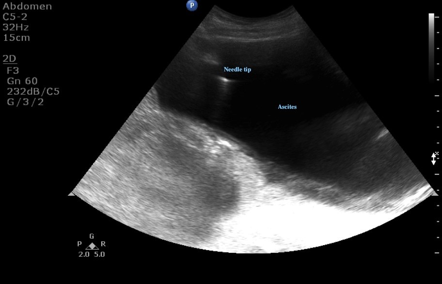
Images brought to us by Drs. Jann Blanton and Miriam Fischer. They used real-time ultrasound guidance for performing a paracentesis. The first image was obtained in the right upper quadrant with the indicator towards the patient's head.


Here we clearly see free fluid in Morrison's pouch that extends superiorly around the liver. Next, the probe is moved to the lower abdomen to identify the largest pocket of free fluid. In the second image, we see the needle entering the peritoneal cavity obliquely from just beneath the indicator marker.
Sometimes ascites is easy to identify with physical exam alone, but other times the exam isn't as clear. Ultrasound can help us to confirm the presence of ascites, and ensure that there is sufficient fluid to safely tap without injury to the surrounding bowel. Ultrasound can also be used to identify the inferior epigastric arteries and the bladder to ensure that these are not in the path of our needle. You can mark the best spot prior to the procedure, or as seen here use the ultrasound in real time. Don't forget to keep your probe sterile if you are going to use it for real-time guidance.
Date: September 2011

