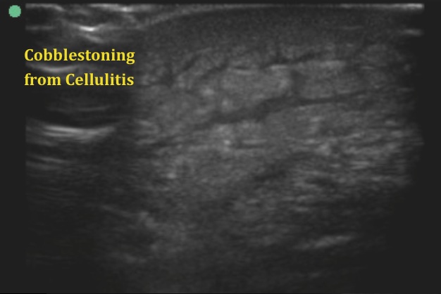
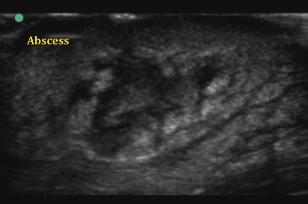
This week's images come to us from Ezaldeen Numur. He used ultrasound to evaluate a patient with soft tissue infection to determine the need for incision and drainage. Ultrasound has been shown in studies to improve the accuracy of detection of abscess compared to exam alone. To do the exam use the high-frequency linear probe. Since we are placing the probe over infected tissue, cover the probe, and clean it well after use. Image 1 shows the classic appearance of cellulitis. The subcutaneous tissues take on a "cobblestone" appearance as fluid collects between fat within the dermis. As the infection continues to progress, an abscess can form. Seen in image 2, abscesses are hypoechoic relative to surrounding tissues. The contents are not uniform in appearance, and debris within the cavity on ultrasound will appear to have mixed echogenicity. Some abscesses will have "sonographic fluctuance." When you press gently on the tissue with the probe, debris within the cavity will swirl around.
Ultrasound can improve the safety of your I&D by helping you to identify underlying vasculature. You may also identify the deep extension of an abscess that may not be appropriate for bedside I&D. Make sure to scan through the affected area in two planes to ensure you have seen the full extent of the abscess.
Image 1
 .
.
Image 2
 .
.
Date: March 2013

