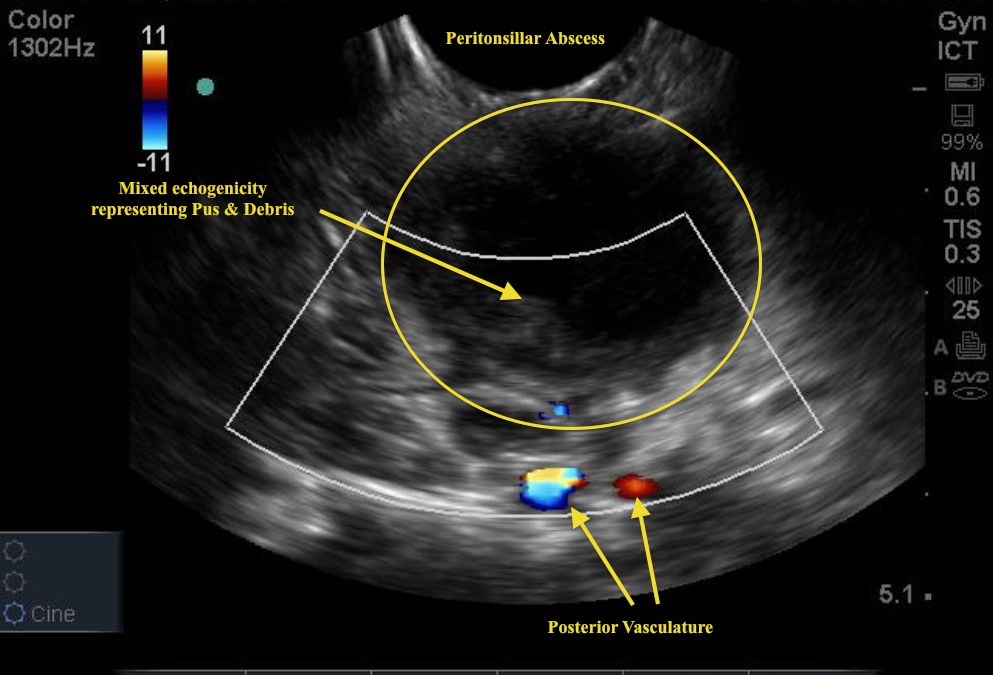
Images come to us from several providers: Drs. Natasha Dehmeri, Deepa Patel, Doug Chesson, and Jeremy Hess. The image is a classic of the diagnosis.
Image 1

Can you identify the pathology? Note the use of the intracavitary probe (exaggerated curve at the top of the screen). The cavity being evaluated in this case is the mouth. Why? To get a better view of what could be causing sore throat, fever, and swelling in the posterior oropharynx. The US is a great tool to confirm the presence of a peritonsillar abscess (PTA) - the diagnosis in this image.
To acquire this view, first place topical anesthesia over the area of interest. Then place a cleaned intracavitary probe with probe cover, orally over the area of swelling. On the US screen, look for an area of the fluid collection - this will likely be a defined area of mixed echogenicity (Image 1). In this image, you see a large PTA. Note the use of color flow to identify the depth of the posterior vessels.
This is a great example of bedside ultrasound confirming a diagnosis as well as helping guide the ED treatment.
Date: November 2012

