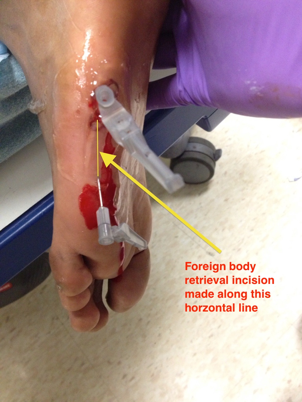
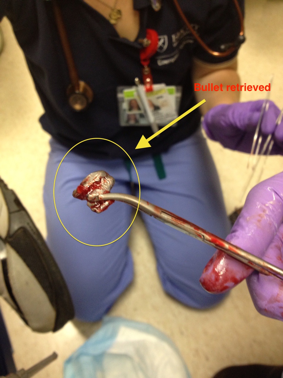
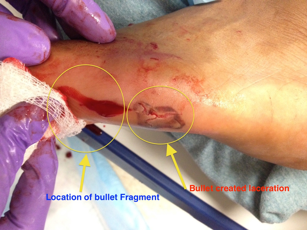
This image shows how ultrasound can be effective in locating and retrieving a foreign body.
Image 1

Image 2

Image 3

How: The patient in this case "stepped" on “something” and presented with a plantar foot laceration. ED XR revealed a 2x1 cm bullet lodged in the foot. The physical exam did not allow for easy discernment of the bullet track and ideal location for incision and removal. However, on ultrasound, the foreign body appeared as a hyperechoic (bright) structure underneath soft tissue, with posterior acoustic shadowing. Dr. Penn then anesthetized the plantar foot by performing an ultrasound-guided posterior tibial and a landmark guided sural nerve block. Subsequently, he inserted an 18-gauge needle in-plane with the ultrasound probe, under dynamic ultrasound visualization (in real-time while looking at the ultrasound screen) on each end of the foreign body. He then made a linear incision using a scalpel along the path created by the two needles. Then after only about a minute of careful searching using a hemostat, the foreign body was successfully retrieved.
Clinical Importance: The foreign body was not easily palpated, but required removal because of plantar foot location. Without ultrasound, this procedure would have been laborious and unnecessarily painful.
Literature Support: Dean AJ, Gronczewski CA, Costantino TG. The technique for emergency medicine bedside ultrasound identification of a radiolucent foreign body. J Emerg Med 2003;24(3):303-8.
Date: August 2013
Image credit: Who: Dr. Brandon Penn.

