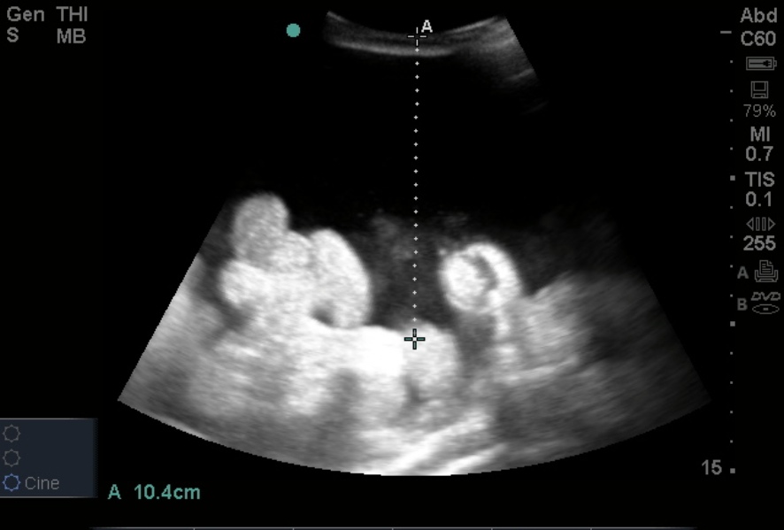
While performing a US assisted paracentesis, the inferior epigastric vessels were identified.
Note the color flow and pulsatile nature of the vessels with a small amount of free fluid surrounding small bowel. Injury to the epigastric vessels resulting in hematoma formation, persistent bleeding, and/or bloody tap are known complications of abdominal paracentesis. Bedside US is essential in confirming the clinical suspicion of ascites, identifying safe deep fluid-filled pockets for the tap, AND may also identify and help you avoid the epigastric vessels.
Prior to performing the procedure, pre-scan your patient with the curvilinear probe looking for deep pockets of fluid (Image 1). Note how the bowel floats in the hypoechoic free fluid. The deepest pockets are often in the flanks, well away from the common anatomical position of the inferior epigastric vessels.
Once you have confirmed ascites, find your desired location for the procedure. Most providers will perform the procedure in static fashion rather than with direct US guidance/visualization of the needle into the peritoneum. If entering more medially in the abdomen, strongly consider localizing the epigastric vessels with US to avoid this potential complication (Image 2). You can use the curvilinear probe to find the vessels. However, the linear high-frequency probe (as used in the video) is best for superficial structures.

Date: May 2012

