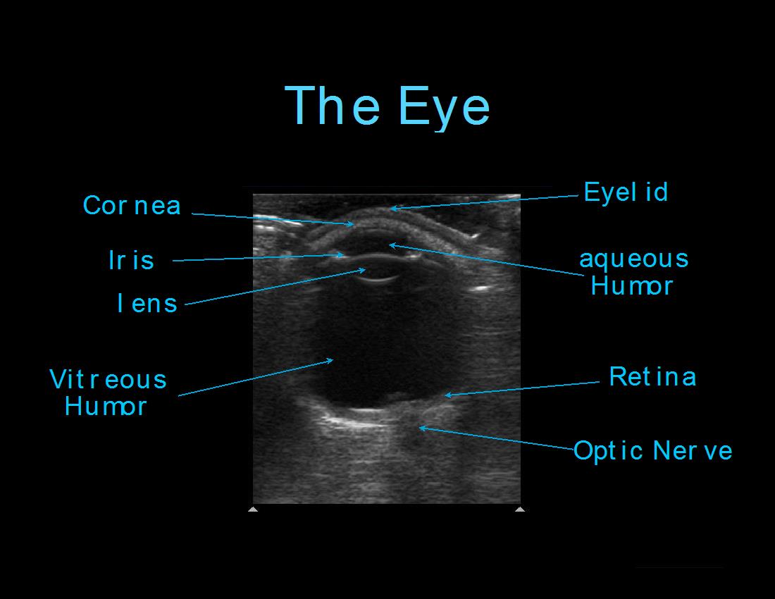
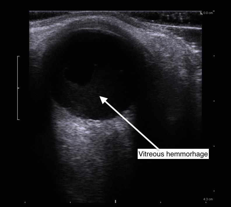
This week’s Image comes from Drs Tibaleka and Pomerleau, who were working in the Marcus Trauma Unit when they encountered a 60-year-old male who reported he had gasoline splashed into his eye with painless vision loss 2 weeks prior. They performed a bedside ocular scan which showed echogenic material in the posterior chamber of the eye. Vitreous hemorrhage was suspected and the patient received an urgent Ophthalmology consult.
Image 1

Image 2

The eye is an ideal organ to scan – thanks to its great acoustic properties – it is largely fluid-filled. The ultrasound appearance of a vitreous hemorrhage depends on its severity and age. Fresh mild hemorrhage may appear as punctate or linear areas of hyperechogenicity. Older or more severe bleeding in the posterior chamber will appear organized. Thrombus can have the appearance of swirling with the extra-ocular movement of the globe.
A quick review of the technique to obtain ocular ultrasound:
Apply a transparent dressing to the closed eyelids of each eye of the patient and apply copious amounts of ultrasound gel on top of that. Next, using a high-frequency linear transducer, place the probe on top of the mound of gel. Adjust the depth so that the globe fills the majority of the screen. Scan both eyes in transverse and sagittal planes. Limit the time of exposure the eye receives to ultrasound since theoretically there could be adverse risks to ocular tissue as the sound is converted to heat energy in the eye.
February 2015

