Ocular ultrasound patients
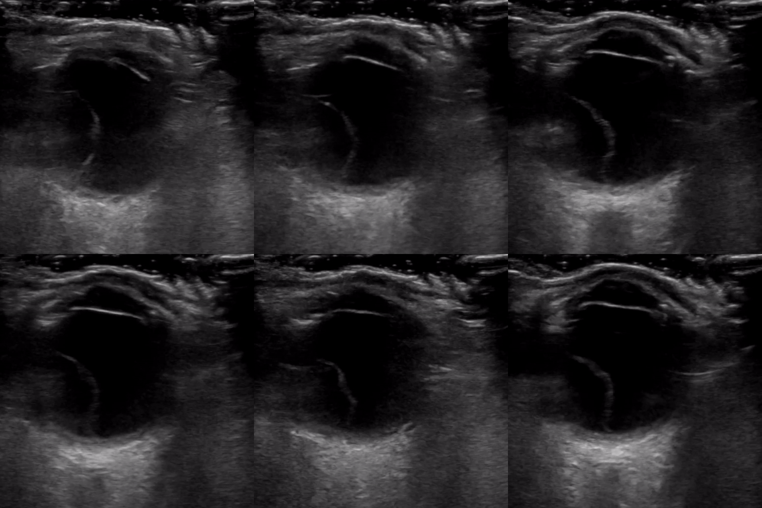
The first patient was a previously healthy elderly gentleman who presented to hospital reporting unilateral, central vision loss and floaters for a week. Vitals signs and 10-point ROS were otherwise normal. Bedside ultrasound is shown below:

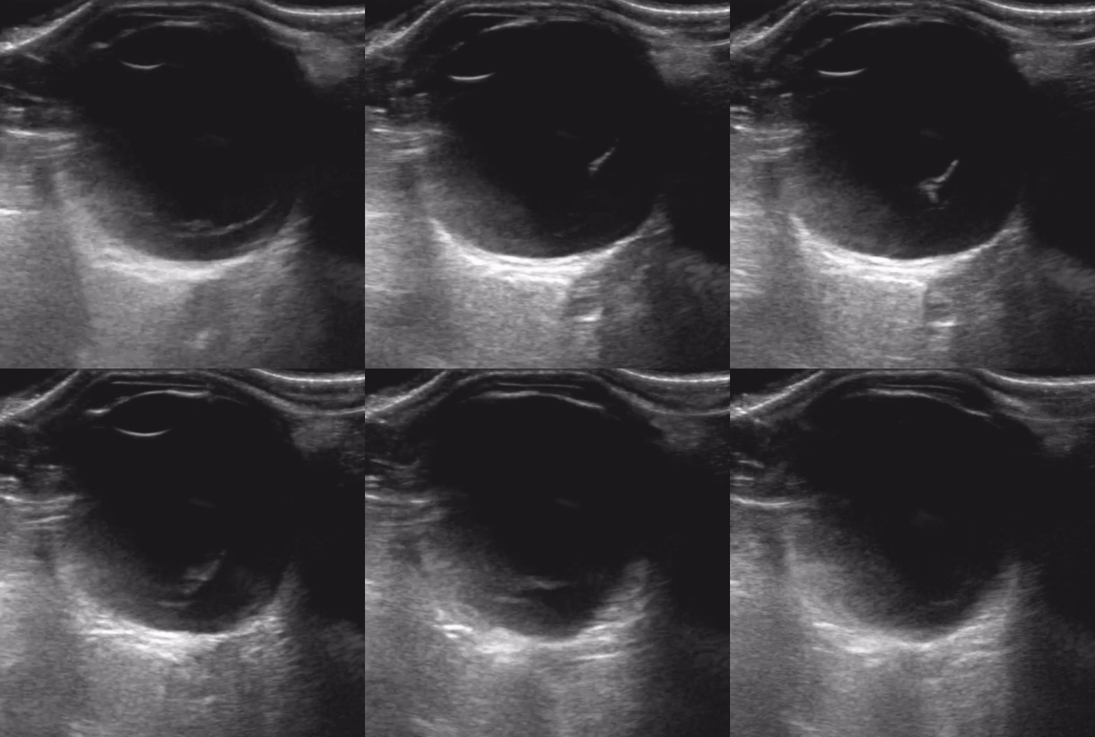
Compare this image with another ocular ultrasound performed in the ED that week in a middle-aged woman who presented with 2 days of floaters and unilateral central vision loss. She too had normal vital signs and normal external ocular exam.

These were similar clinical presentations, but in the first ultrasound, there is a mobile flap just anterior to the retina that is tethered to the optic nerve posteriorly and ora serrata anteriorly [arrows]. This is typical of a retinal detachment. The second video demonstrates a vitreous detachment, where an undulating membrane is seen beyond the limits of the optic nerve, and that continues to move after eye movement has stopped.
Ultrasound is a great tool to distinguish the two, especially because retinal detachment requires immediate ophthalmologic referral to prevent permanent vision loss. Patients with vitreous detachment also require close follow-up with ophthalmology, but less urgency than retinal detachment. It is important to sweep through the globe in two planes thoroughly to avoid misidentifying a retinal detachment as a vitreous detachment - if in doubt, err on the more conservative side, and request urgent ophthalmology consult.
Authors: Dr. Jasmin Tanaja, and Dr. Layne Madden

