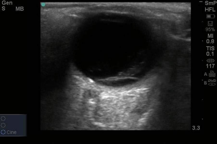
This week's IOW is brought to us by Sultan Alwajeeh, EM PGY-1 and Dr. Kyle Minor - this pt was referred from outside Grady due to a concerning exam.
Look at this video link first.
What are the two ED diagnoses that come to mind with this video?
1) Retinal Detachment
2) Vitreous Hemorrhage
To acquire this image, lay pt supine and have eyes looking up in neutral position. Use sterile jelly and place it over the closed eyelid. Some may prefer to place a tegaderm over the closed eye first and then generously place sterile gel (single use KY packet is ok) and place the probe over the eye in transverse fashion. With gentle contact, fan the high-frequancy linear probe over the eye lid. Subtle movements should bring the anatomy into view. You should try to find the location of the optic nerve as most detachments are still anchored by the optic nerve. You may wish to have the pt move the eye while the lid is closed - this may help you better see the pathology.
The below image labels the pathology - a retinal detachment. Notice how the retina - a structure that should be intimately attached to the posterior surface of the eye (choroid) - has pulled off. The patient had acute visual symptoms and was referred emergently to Opthalmology based off of this exam.
For more reading on bedside US in the ER, see the linked article from Academic Emergency Medicine, 2000.
6/4/12


