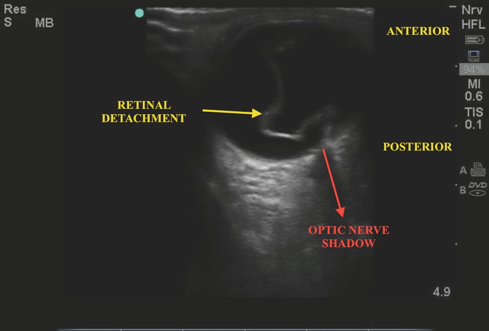
What is the ED diagnosis based on the link below?
To evaluate the eye with ultrasound, lay the patient supine with the eyes looking up in a neutral position. Use sterile jelly and place it over the closed eyelid. Some may prefer to place a Tegaderm over the closed eye first and then generously place sterile gel (single-use KY packet is ok) and place the probe over the eye in a transverse fashion. With a gentle contact, fan the high-frequency linear probe over the eyelid. Subtle movements should bring anatomy into view. You should try to find the location of the optic nerve, as most detachments are still anchored by the optic nerve. You may wish to have the pt move the eye while the lid is closed - this may help you better see the pathology.
The below image labels the pathology - a retinal detachment. Notice how the retina - a structure that should be intimately attached to the posterior surface of the eye - has pulled off. Common presenting symptoms include "flashing lights" and "floaters" in the visual field. Retinal detachments are classically painless and are more commonly seen in patients with a history of cataract surgery, diabetes, trauma, connective tissue disorders, or with a family history of retinal detachment. Although fundoscopy is another means to the diagnosis, the findings are much less obvious than those generally seen with ultrasound.
Image 1

Date: November 2012
Image credit: Carmelle Tabuteau and Justin Schrager

