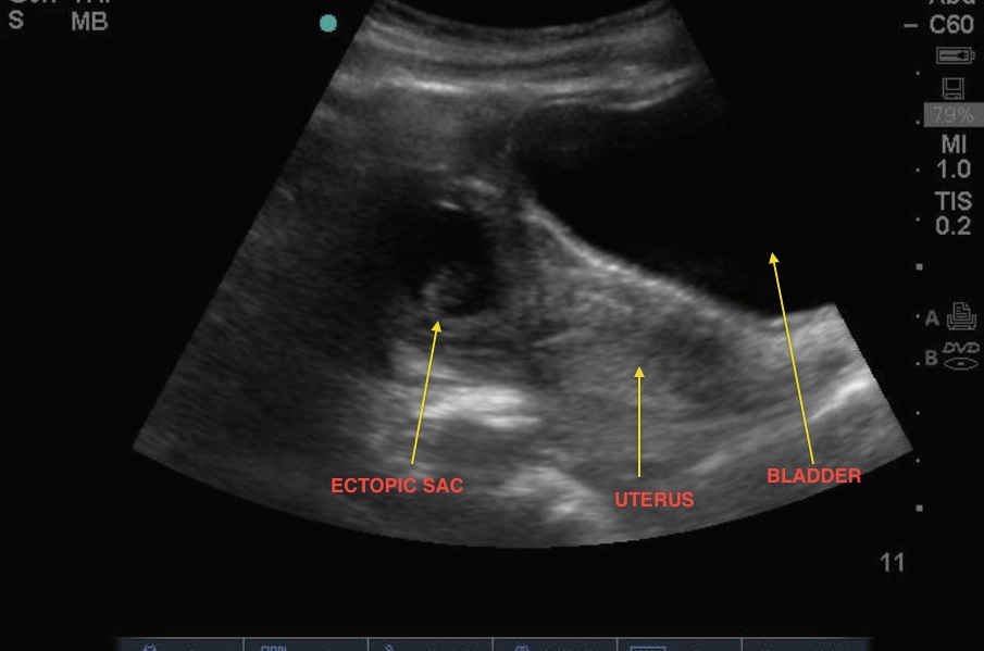
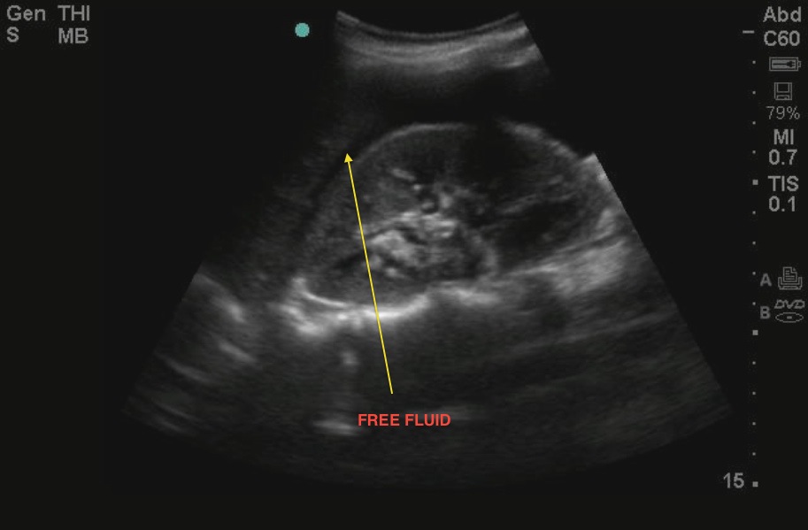
This patient presented hypotensive with severe abdominal pain. The pregnancy test was positive. ED bedside ultrasound revealed a positive FAST; anechoic free fluid was noted in the RUQ and in the pelvic views. On pelvic transabdominal scanning, a gestational sac was noted with fetal pole measuring 9 weeks and positive fetal cardiac activity. In certain views, the pregnancy appeared to be within the uterus, but with fanning, the abnormal location was correctly identified at the bedside and the patient went emergently to the OR with OBGYN. This case emphasizes the importance of careful scanning in cases of suspected ectopic pregnancy. It is important to scan in both the transverse and longitudinal planes and fan the entire uterus to fully assess gestational sac implantation location. The thinnest wall of the uterus surrounding the gestational sac should be at least 8mm in diameter.
Image 1

Image 2

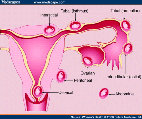
Image 3

Clinical Importance
An interstitial ectopic pregnancy (often also called a cornual ectopic) is a type of ectopic pregnancy. It accounts for 2 - 4% of all ectopics. It occurs when the pregnancy implants in the interstitial portion of the fallopian tube (see image). These pregnancies can easily be misidentified as normal intrauterine pregnancies on ultrasound. They tend to rupture later (8-12 weeks) and bleed heavily when ruptured. The morbidity and mortality are higher due to later presentation and associated complications.
Literature Support
Moawad NS, Mahajan ST, Moniz MH, Taylor SE, Hurd WW. Current diagnosis and treatment of interstitial pregnancy. Am. J. Obstet. Gynecol. 2010; 202: 15–29.
Date: October 2013
Image credit: Drs. Stokes and Blanton

