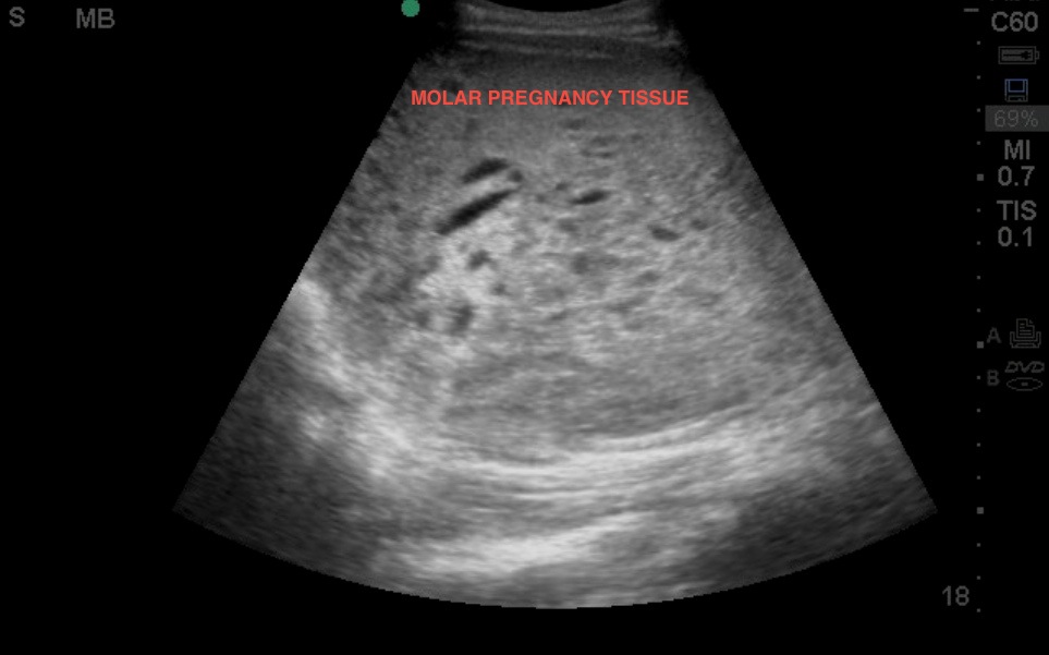
In symptomatic pregnant patients, the goal of point of care ultrasound is to identify a definitive intrauterine pregnancy (IUP). A definitive IUP is defined as a gestational sac containing a fetal pole or yolk sac. This exam begins with transabdominal imaging of the uterus using the curvilinear probe. The uterus is imaged in sagittal (indicator marker to toward the patient's head) and transverse planes (indicator marker towards the patent's right). Typically the endometrial stripe can be easily identified. In this image, no IUP is identified and the endometrial stripe is obliterated by a large mass with solid and cystic components. This was subsequently diagnosed as a complete molar pregnancy. Ultrasound findings in molar pregnancy can vary from the classical image of a “snowstorm” pattern seen when using older ultrasound technology, to the complex mass of swollen hydropic trophoblastic tissue as seen here with higher-resolution equipment.
Image 1

Clinical Importance
Hydatidiform molar pregnancies may be complete or partial, depending on their chromosomal complement. Complete moles (46 XX, or 46 XY) contain no fetal tissue,. However, partial moles (69 XXX, or 69 XXY) often have fetal tissue present. Both develop from an aberrant fertilization event that leads to the development of an abnormal proliferative process. The two main risk factors for the development of this entity are advanced maternal age and a history of previous gestational trophoblastic disease. Patients may complain of vaginal bleeding or pelvic pressure or pain from the enlarging uterus, which grows much larger than expected for gestational dates.
Literature Support
Saul T, Sonson J. “Molar Pregnancy.” Visual Diagnosis in Emergency Medicine, 2008.
Date: October 2013
Image credit: Drs. Chesson and Shayne

