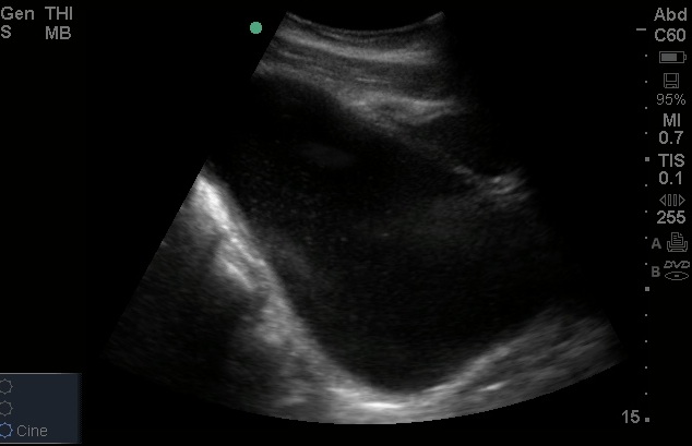
This IOW comes from Dr. Stephanie Cohen, Pediatric EM Attending at Egleston. Can you identify the anatomy in the unlabeled still image (Image 1)?
Image 1

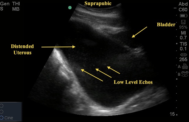
This is the suprapubic view of a 14 yo who female who presented with abdominal pain. Bedside US was performed and revealed findings of a severely distended UTERUS. Note the hypoechoic fluid with some low level echos inside the uterus (Image 2).
Image 2

This is suggestive of accumulated menstrual blood. The findings prompted a complete GU-exam that revealed an imperforate hymen.
Diagnosis: Hematometrocolpos. This is not a common ED diagnosis but develops in the setting of a vaginal obstruction - in this case the imperforate hymen. Your suspicion for this diagnosis should increase in a child with features of puberty, primary amenorrhea, and intermittent abdominal pain. The classic physical exam finding is a bluish bulging membrane at the introitus. US is used to distinguish between hematocolpos (fluid in vagina) vs hematometrocolpos (fluid in BOTH the vagina and the uterus). Treatment is generally an incision of the bulging tissues.
See below for YouTube video links to both the long and sort axis views of the uterus and bladder.
7/26/12

