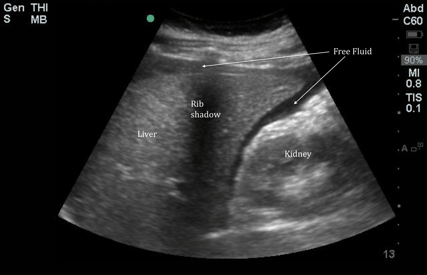
The IOW this week comes from Dr. Patrick Meloy, who used bedside ultrasound to rapidly triage and diagnose a woman who presented to the ER for abdominal pain. Recall that the first view to obtain in a young female patient with a positive pregnancy test and clinical suspicion for a ruptured ectopic is a RUQ FAST view. The presence of free fluid in the RUQ has been show to predict the need for operative treatment in this patient population. In Image 1 note the large quantity of hypoechoic free fluid in Morrison’s pouch and surrounding the liver.
Image 1

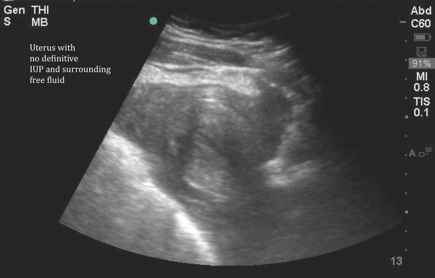
After visualizing the right upper quadrant, an image of the pelvis should be obtained. Image 2 is a sagittal cross-section of the uterus obtained by placing the probe just above the pubic bone with the indicator marker pointed towards the patient's head. In this view, we again see large free fluid. Here the blood around the uterus is partially clotted, so appears complex with internal echoes. In this view, we are also looking for evidence of an intrauterine pregnancy. Here we see the endometrial stripe, but no evidence of an IUP (defined as a gestational sac + either a yolk sac or a fetal pole). The presence of free fluid and the absence of an IUP in a patient with a positive pregnancy test should be considered a ruptured ectopic pregnancy until proven otherwise. In this case, OB-GYN was consulted immediately based on the bedside scan, and pt was taken to the OR.
Image 2
Date: December 2014


