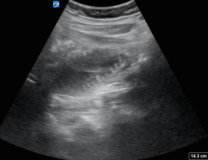
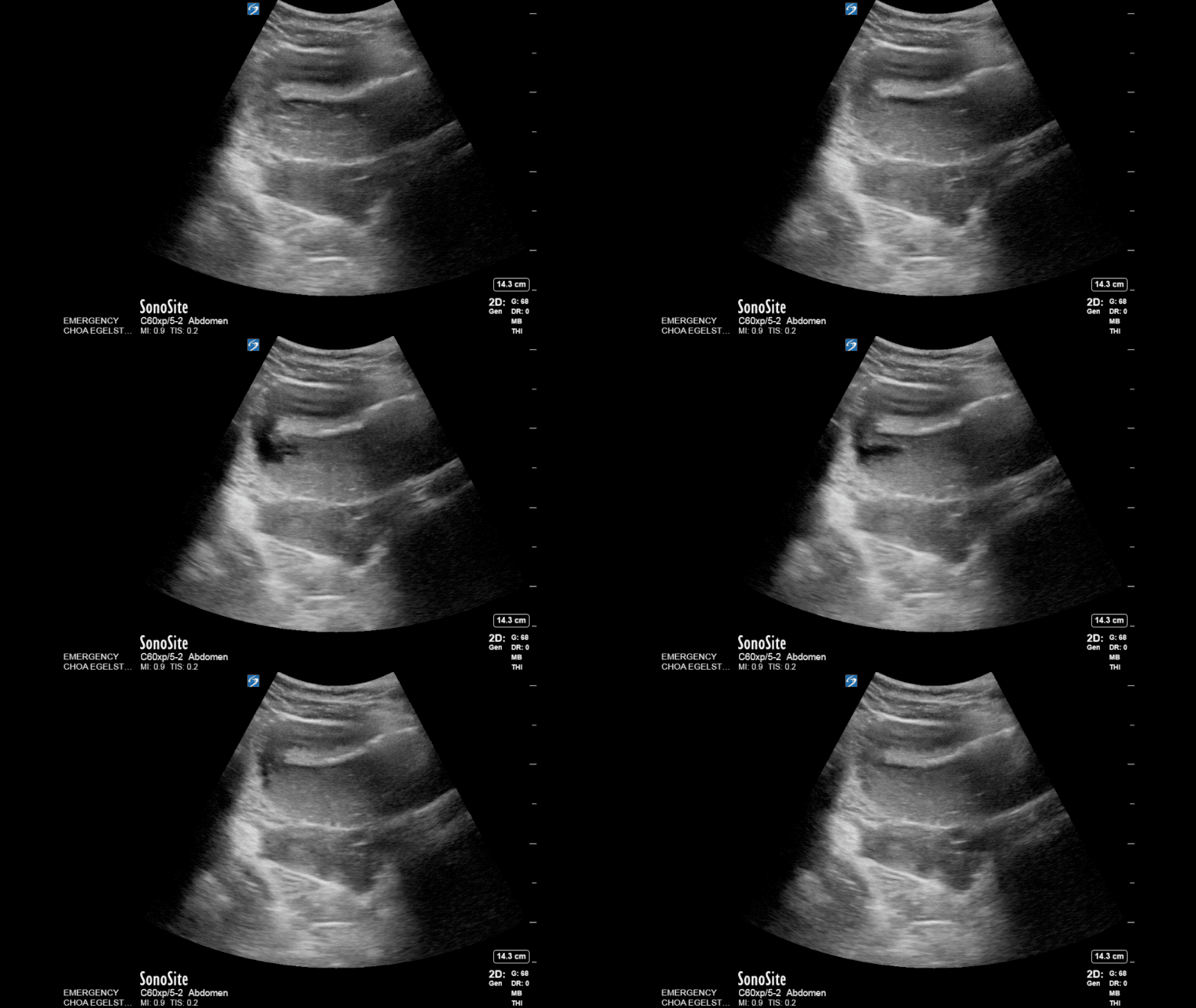
This week’s image comes from Drs. Stephanie Cohen, Colleen Gutman, and Anita Bharath at Egleston Children’s Hospital. The patient is a 12 yo girl with VACTERL (a syndrome characterized by Vertebral, Anal, Cardiac, Tracheoesophageal, Limb abnormalities) and multiple prior abdominal surgeries presenting now with 3 days of emesis and abdominal pain. Emesis was bilious and she had no passage of stool for 2 days. Images obtained are shown below.

Note the visible Plicae circularis of the small intestine. This image is also referred to as the keyboard sign.

Thickened bowel loops and to-and-fro peristalsis are other findings suggestive of a small bowel obstruction.
Ultrasound has better sensitivity and specificity than a plain x-ray for the diagnosis of small bowel obstruction. One metanalysis looked at x-ray, CT, MRI, and US for diagnosis of SBO. SBO was diagnosed if there were >2.5cm loops of small bowel proximal to collapsed bowel segments, and/or with absent or decreased peristaltic activity (defined as to and fro peristalsis). Two Studies looked at bedside ultrasound by emergency physicians in particular. Sensitivity and specificity were 97 and 90% respectively with likelihood ratios of positive 9.5 and negative of 0.04, better than both CT and MRI as well.1,2 Keyboard sign or visualization of the plicae circularis clearly, and to and fro peristalsis are other features of SBO.3
The patient was admitted to the surgery service, and after a period of observation in the hospital was taken to the OR and had a partial small bowel resection and lysis of adhesions.
References
- Taylor MR and Lalani N. Adult Small Bowel Obstruction. Acad Emerg Med. 2013; 20(6):528-44.
- Jang TB, Schindler D, and Kaji AH. Bedside ultrasonography for the detection of small bowel obstruction in the emergency department. Emerg Med J. 2011; Aug;28(8):676-8.

