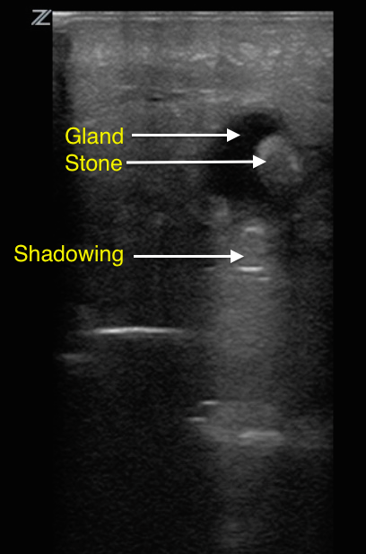
The linear probe is best used to image superficial structures as it provides rich detail. In case of unilateral or painful swelling in the region of the submandibular or parotid gland, place the linear probe over the cheek/face and attempt to identify an obstructing stone and dilated associated salivary gland. Salivary stones appear as hyperechoic lines with posterior acoustic shadowing within a dilated salivary duct. Stones smaller than 2-3 mm are difficult to identify due to a lack of acoustic shadowing1.
Image 1

Clinical Importance
Sialolithiasis with salivary gland obstruction is a rare disease and can mimic more frequently occurring illnesses such as facial and dental infection or abscess. Salivary stones located in the gland or duct system can be diagnosed using high-frequency sonography, and these findings can be differentiated from the ultrasound appearance of cellulitis and abscess2. Additional information may be obtained by sonography such as number, size, and location of stones to determine the prognosis of stone passage and may guide initial management of the symptomatic patient in the emergency setting3.
Literature Support
- Emergency Medicine, An Issue of Ultrasound Clinics - By Mike Blaivas
- Duprey et al. Emergency Physician point-of-care ultrasound in the diagnosis of sialolithiasis
- Hoffman, B. Case Report - Sonographic bedside detection of sialolithiasis with submandibular gland obstruction

