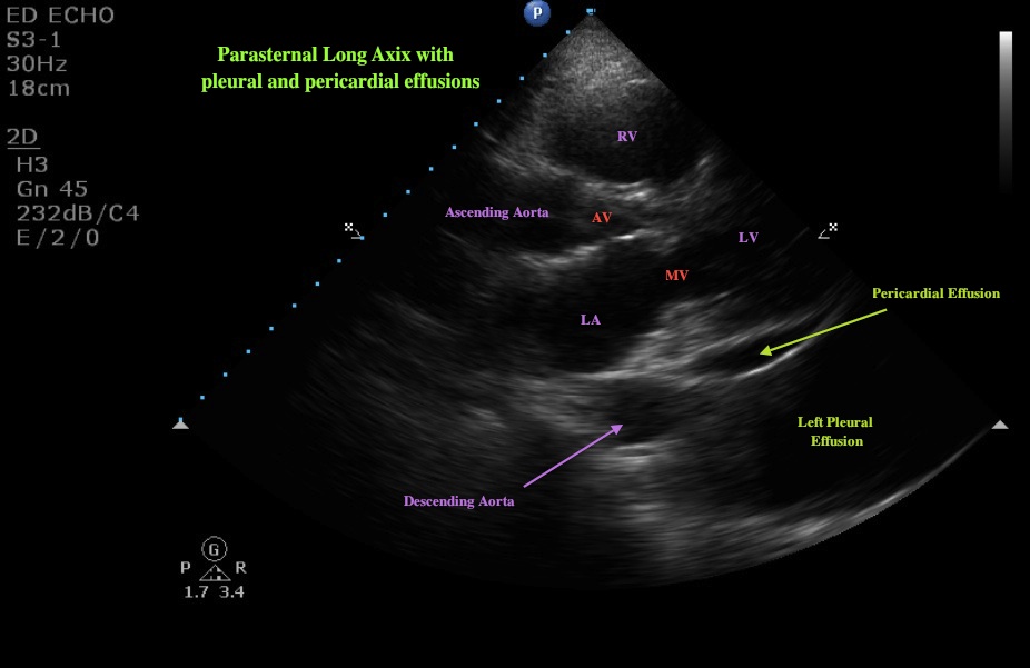
This week’s image is brought to us by Drs. Patrick Thomas and Scott Kurpiel. It is a parasternal long-axis image with evidence of both a pericardial effusion and a left pleural effusion.
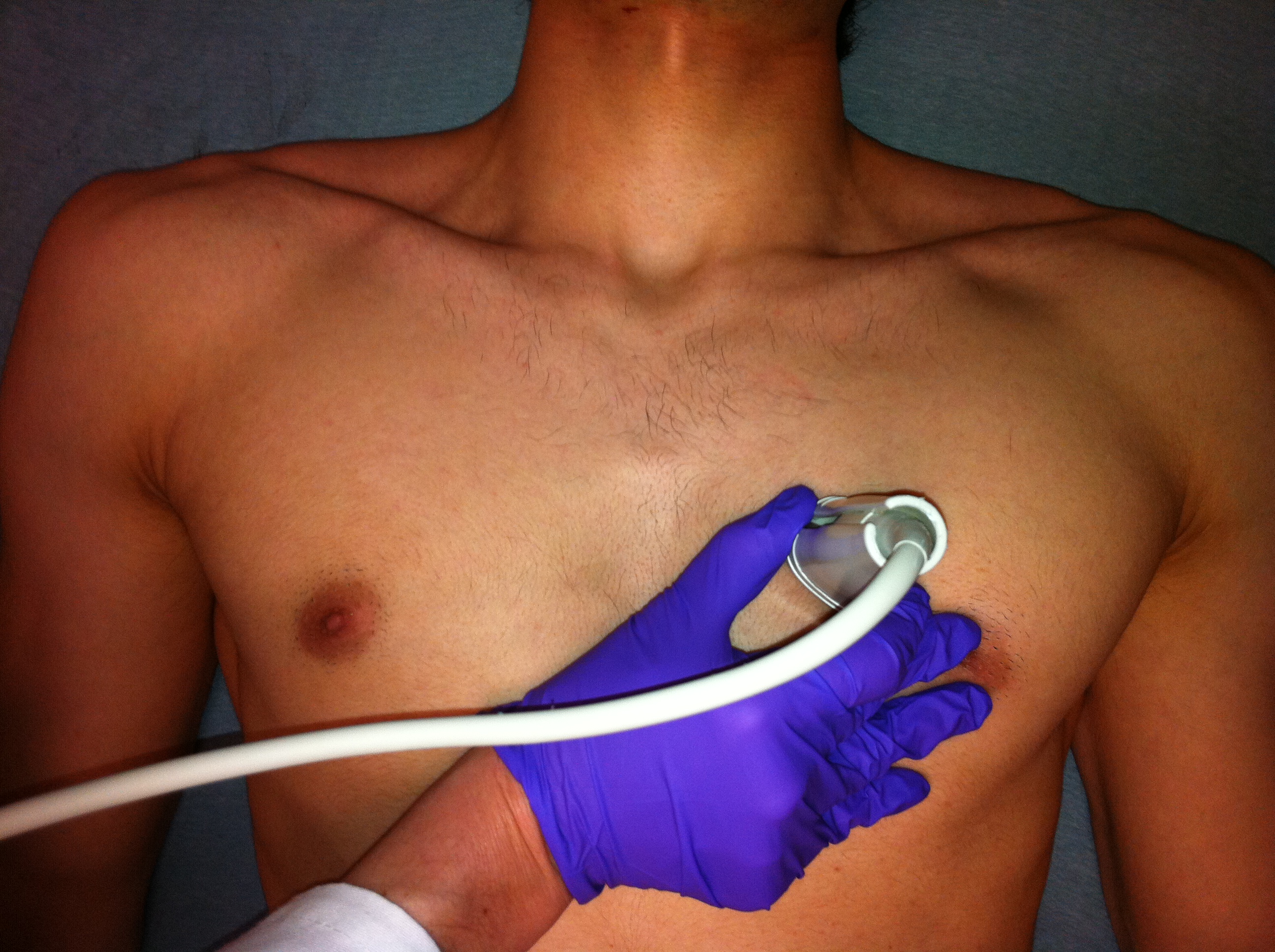
The view is obtained by placing the probe at the left sternal border in the 3rd or 4th intercostal space. The indicator is pointed toward the patient’s right shoulder. The ideal image in this axis will show us a cross-section of both the mitral valve and the aortic valve. Achieving this may require some slight fanning or rotation of the probe.
This still image is taken in diastole. The MV leaflets are open and the septal leaflet is slapping the septum (a marker of good LV function). Beneath the heart, we see the descending aorta in cross-section. This is a key landmark for distinguishing a left pleural effusion from pericardial effusion. The pericardium tracks anterior to the descending aorta and thus a pericardial effusion will do the same. Pleural effusion will track distal to the aorta as seen in this image.


Date: October 2011

