The image of the week is a great video brought to us by Drs. Kyle Brown and Adam Pomerleau.
They used the US to correctly diagnose a pneumothorax by finding the "Lung Point," the only ultrasonographic finding that is highly specific for pneumothorax. Recall that as part of our E-FAST exam we look for lung sliding in the second or third intercostal space, roughly mid-clavicular line. With the indicator towards the head, the pleura will appear as a bright hyperechoic line flanked on both sides by a rib with shadowing beneath. We use the presence of lung sliding to rule out pneumothorax.
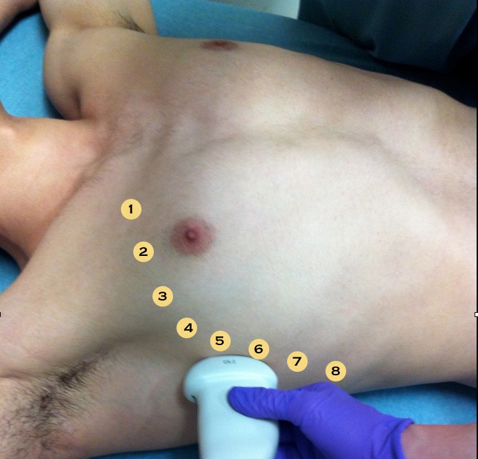
If we don't see sliding we can continue our evaluation by moving the probe evaluating each rib space down and out laterally (see image). The lung point first described by Lichtenstein is the appearance of a lung pattern replacing a pneumothorax pattern at a single location on the chest wall. As the patient breathes and the lung expands, it will move in and out of view underneath your probe. On the left chest, it is especially important that you move lateral enough as the beating of the heart can be a common fake-out for a lung point.
Remember that the lung point is a highly specific finding but has low sensitivity. If the lung is completely collapsed you will never find the lung point because the lung is too small and never comes into contact with the chest wall.


