Place the linear probe on the chest oriented in the longitudinal direction with the probe marker pointing to the patient’s head. On ultrasound, ribs appear hyperechoic (bright) with shadowing posterior. Pleura is seen as a hyperechoic straight line extending between two rib shadows. Optimal screen depth allows for two ribs to flank the screen with pleura between. There is a shimmering motion noted at the pleural interface known as pleural sliding. Normal healthy lung without pleural injury will exhibit pleural sliding.
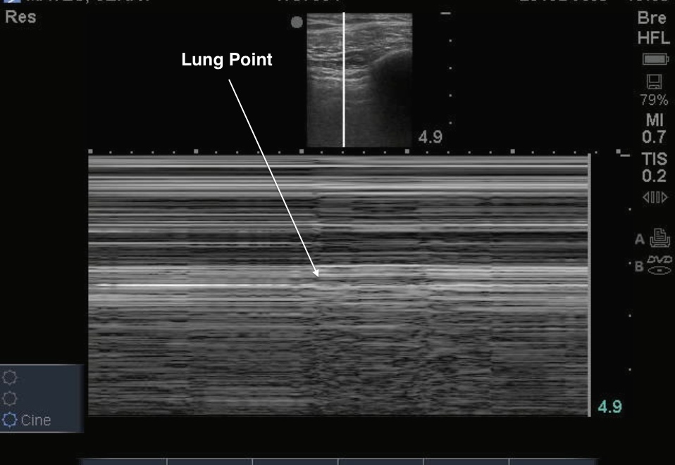
The interface of where healthy lung starts and where the pneumothorax ends is known as the lung point. On one side of the lung point, healthy pleura will be seen with pleural sliding, whereas on the other the pneumothorax will show a still pleural line with absent sliding.
Image 1

Clinical Importance: As larger pneumothoraces are more likely to require thoracostomy, it is important to determine the size of the pneumothorax. The location of the lung point may assist in determining the size of the pneumothorax. If a lack of lung sliding is visualized anteriorly, the probe can progressively be moved to more lateral and posterior positions on the chest wall searching for the location of the lung-point. The more lateral or posterior the ‘lung-point sign’ is identified, the larger the pneumothorax1. Therefore, if the ‘lung-point sign’ is seen in an anterior location on the chest wall, the pneumothorax is relatively small. The lung point sign had an overall sensitivity of 66% and a specificity of 100%2. Keep in mind that the sensitivity may be low because in large pneumothoraces, the lungs are so collapsed that there is no point when the inflated lung is in contact with the parietal pleura.
Literature Support:
- Husain L, Hagopian L, Wayman D, Baker W, and Carmody K. Sonographic diagnosis of pneumothorax. J Emerg Trauma Shock. 2012 Jan-Mar; 5(1): 76–81.
- Chan S. Emergency Bedside Ultrasound to Detect Pneumothorax. Acad Emerg Med January 2003.

