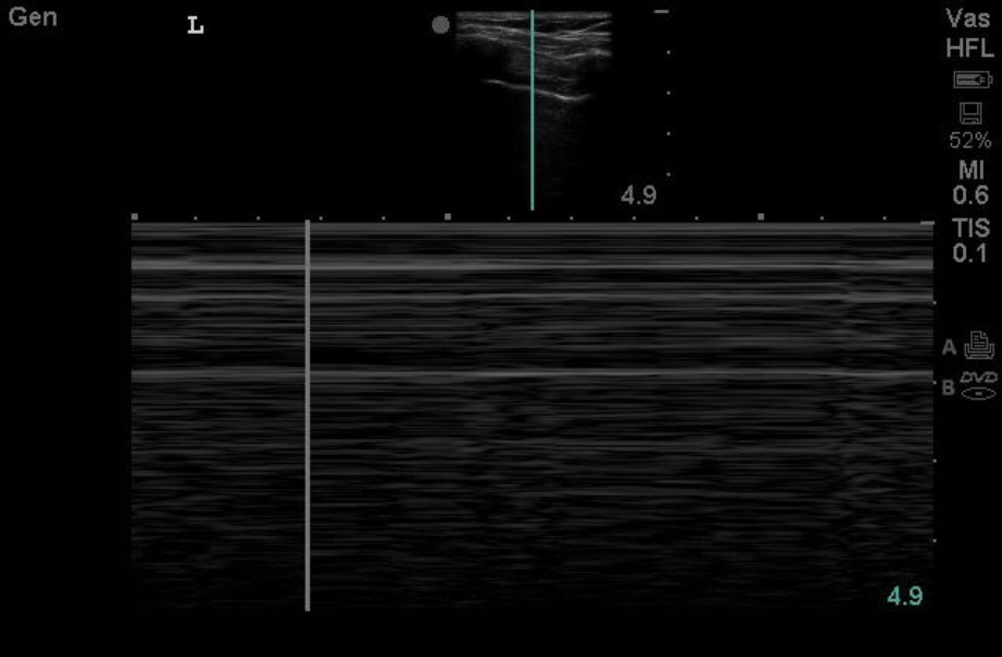
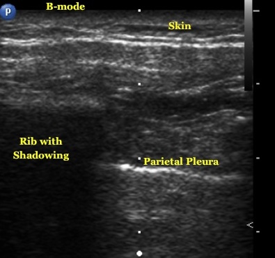
Place the linear probe on the chest oriented in the longitudinal direction with the probe marker pointing to the patient’s head. On ultrasound, ribs appear hyperechoic (bright) with shadowing posterior. Pleura is seen as a hyperechoic straight line extending between two rib shadows. Optimal screen depth allows for two ribs to flank the screen with pleura between. There is a shimmering motion noted at the pleural interface known as pleural sliding. Normal healthy lung without pleural injury will exhibit pleural sliding.
Routinely ultrasound is performed in B-mode, which is a 2D live representation of the scanned image. The lung sliding sign is noted in B-mode. However, M-Mode may be used to diagnose pleural injury by simply pressing the button labeled M-mode on most ultrasound machines. M-Mode, which stands for motion mode, is a still representation of the B-mode image, with motion represented using still images. Healthy pleura with sliding shows artifact and a difference in motion artifact posterior to the pleura, which is known as the sea shore sign.
Image 1

Image 2

Clinical Importance: Ultrasound is highly sensitive for detecting pneumothorax, much more so than chest plain film imaging. On ultrasound, pneumothorax is detected in B-mode by a lack of pleural sliding and on M-mode by a lack of motion artifact known as the bar code or stratosphere sign.
Literature Support: Blaivas et al. A Prospective Comparison of Supine Chest Radiography and Bedside Ultrasound for the Diagnosis of Traumatic Pneumothorax. Academic Emergency Medicine 2005.
Date: October 2015
Image credit: Todd Taylor

