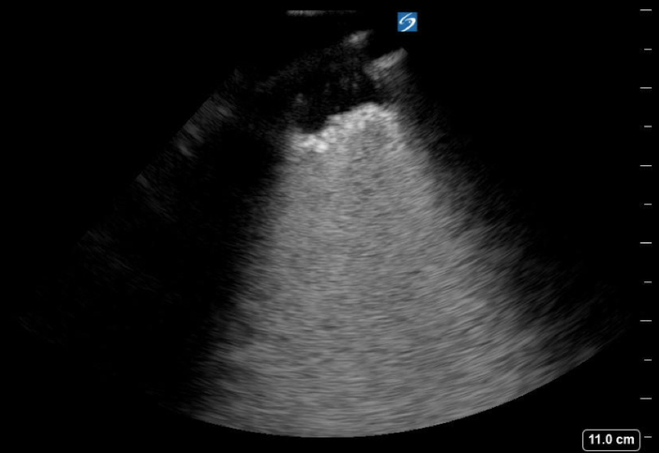
Our image of the week is brought to you in impressive fashion by the ultrasonographic prowess of Drs. Bellerose and Abramoff. Their patient had a history of metastatic malignancy, and presented with fever, cough, and septic shock. Her lung ultrasound showed the following finding. What is that?

Recall that normal aerated lung demonstrates an A-line pattern with horizontal, regularly spaced, bright white, parallel lines generated as a reverberation artifact of the pleura. Instead in this lung ultrasound we see a complete white-out of the rib space with a dark area adjacent to the pleura. This is a C line which indicates lung consolidation. The dark part of the image adjacent to the pleura represents a small focal area of consolidation. Deep to that there is an area of white-out which represents lung that is still aerated but has a very high fluid content. Here's an image to review common patterns of lung artifacts.

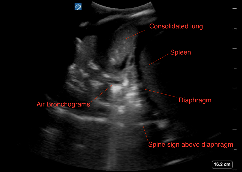
Another image obtained by our esteemed colleagues below further consolidates our diagnosis of pulmonary consolidation (pun intended).

This image was obtained in the left lung base. The far-field region shows a continuation of the spine above the diaphragm indicating pathology in the chest. The pulmonary parenchyma looks similar to the liver or spleen, as there is no longer air in alveoli. Within the consolidated lung we see linear hyperechoic areas which represent sonographic air-bronchograms.
Usama Khalid MD
Ultrasound Fellow
Department of Emergency Medicine
Emory University SOM

