This week’s image is from a middle-aged patient who presented for shortness of breath and lower extremity edema. See if you can find the cause of the patient’s SOB on ultrasound.
Image 1
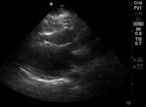
Image 2
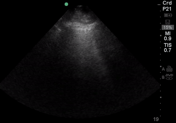
Many of you chimed out in unison: “There are pulmonary B-lines! I can see them in image 2!” You are absolutely correct— the comet-tail artifacts from the lung pleura radiating to the edge of the screen are B. They are certainly concerning for volume overload in a patient with lower extremity edema and SOB; however, the parasternal long-axis view of the patient’s heart (image 1) appears to show a relatively preserved EF. This is why the doctors investigated further with the next still image. Do you know what this image shows?
Image 3
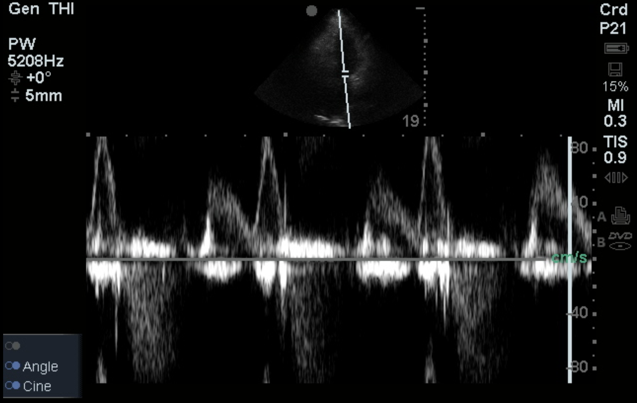
Using the pulse wave doppler setting and setting the doppler gate between the leaflets of the mitral valve on an apical four-chamber cardiac view, were able to assess the patient’s diastolic function and determined that the patient had stage I diastolic dysfunction consistent with impaired relaxation.