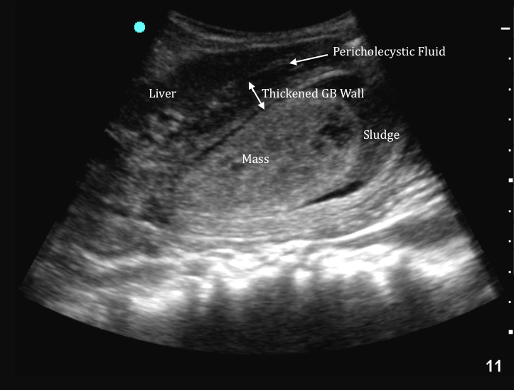
The IOW this week is a markedly abnormal gallbladder from a point-of-care ultrasound. Their patient was a 68 year-old female who was undergoing a workup for undifferentiated altered mental status. The patient denied complaints, but per family was not acting normal. Vital signs were significant for fever and tachycardia. Her initial exam, cxr, & urinalysis did not suggest a source of potential infection. Her labs, however, showed elevated Alkaline phosphatase, AST and ALT, which prompted them to perform a RUQ US. The image below was obtained.
Image 1

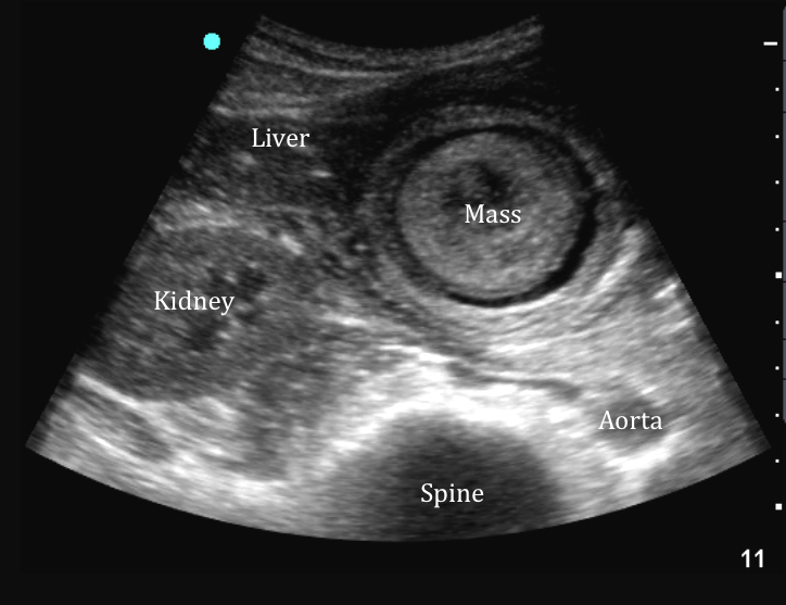
Note the presence of a thickened gallbladder wall and pericholecystic fluid, which suggest a diagnosis of cholecystitis. There is also a large nonshadowing mass in the center of the gallbladder. A polypoid mass within the gall bladder may represent adenocarcinoma, a benign adenoma, polyp, focal adenomyomatosis, metastases, or tumefactive sludge. Tumefactive sludge occurs when sludge fills the gallbladder and take on a solid form that mimics a tumor. Below is a short axis image of the gallbladder.
Image 2

Following the scan, the patient received IV antibiotic directed at the billiary source of infection. Further imaging showed common bile duct stones in addition to the abnormal findings above. She underwent ERCP and cholecystectomy and awaited pathology results on the gallbladder mass.
Date: 2014

