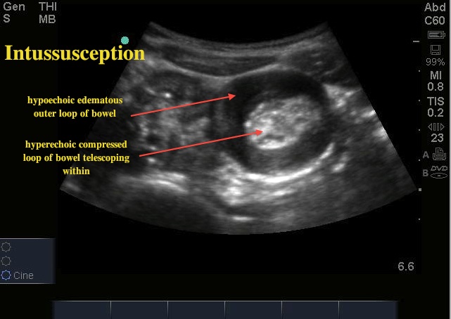
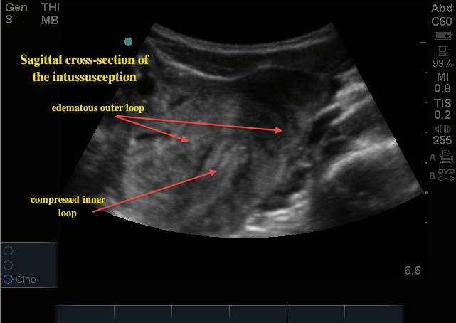
This week's image of the week used bedside ultrasound to make the diagnosis of intussusception.
Those of you studying for the in-service exam will recall that the classic presentation of intussusception is crampy abdominal pain, currant jelly stools, and a sausage-like RUQ mass in a toddler-aged child. This case emphasizes that classic presentations are not so common. This patient was a 7-year-old who presented with colicky abdominal pain. She was found to have a Meckel's diverticulum as the lead point for her intussusception.
The "bulls eye" or "target sign" is the most commonly described ultrasound appearance of an intussusception (see first image). This is an axial cross-section of the affected bowel. There is a thickened hypoechoic rim representing the edematous outer bowel, and a hyperechoic center secondary to the compressed telescoping inner segment. The second image shows the intussusception in sagittal cross-section.


To obtain these images the curvilinear probe was used. In a small child the high-frequency linear probe could also be used. If a mass is palpated, this area should be scanned first. If there is no mass, the entire abdomen can be scanned in two planes in a systematic fashion to find the classic target sign.
2012

