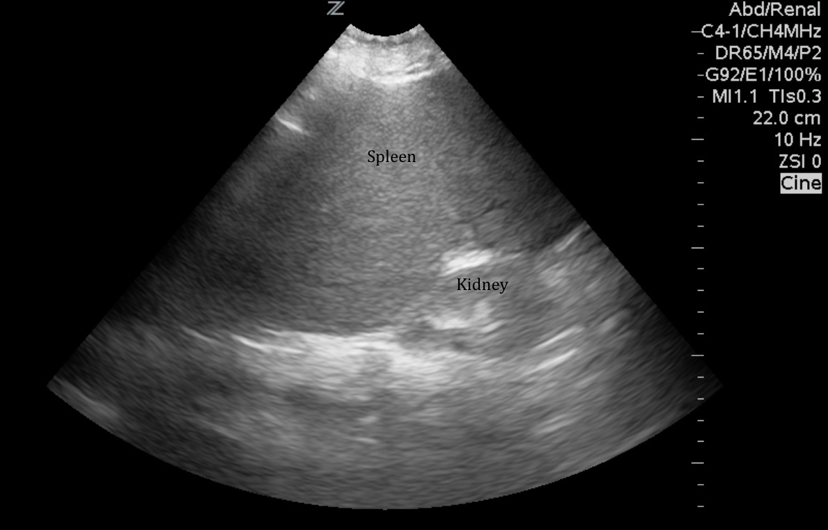
The IOW this week is from a bedside ultrasound to diagnose splenomegaly. To get this image, a curvilinear probe is used to obtain what is essentially a LUQ FAST view. The probe is placed on the patient's left over the lower ribs in the mid axillary line, and the indicator marker is pointed towards the head. Splenomegaly is defined as a spleen that measures >13cm in length in its longest diameter. If rib shadows get in your way, angle the probe to fit between the ribs and make sure to fan towards the back as the spleen is typically more posterior. In general the spleen should be approximately the size of the kidney. Note that in the image below it is significantly larger than the adjacent kidney. Patients with splenomegaly may present with vague LUQ pain. Recall that an enlarged spleen can be seen in infection (CMV, toxoplasmosis, mononucleosis, TB, or malaria), hematologic disorders (myelofibrosis, lymphoma, leukemia, thalassemia), portal congestion (portal htn, portal/splenic vein thrombus, CHF), or other malignancies (hemangioma, mets). This patient presented with LUQ pain and had a diagnosis of acute myeloid leukemia.
Image 1

Sierra Beck MD
Assistant Professor
Department of Emergency Medicine
Emory University SOM
2014

