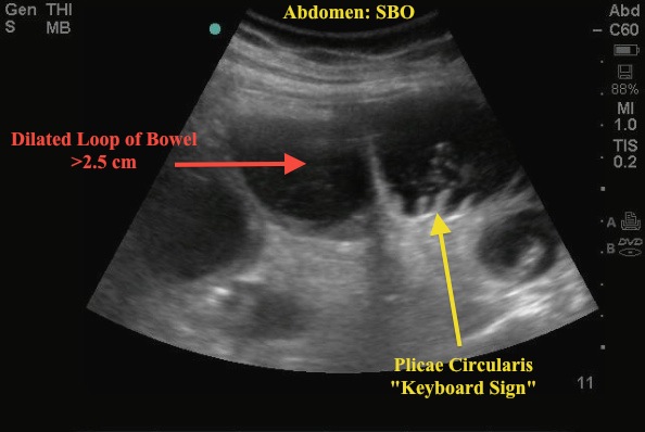
The patient presented with abdominal pain. Bedside ultrasound quickly captured Image 1. Notice a tubular structure, with a mixture of fluid (hypoechoic area) and debris (low-level echos). Also, note the image captures the classic "keyboard" sign (visualization of the plicae circularis) associated with the diagnosis - a small bowel obstruction (SBO).
Image 1

To diagnose SBO with the US, use the curvilinear probe and scan systematically over the abdomen. Look for fluid-filled, dilated loops of bowel (defined as >2.5cm). You may also see back and forth movements of echoes within the lumen as bowel contents move with dysfunctional peristalsis. The plicae circulares can be prominent as seen in this image. Although history, physical exam, and XR findings are the classic method to diagnose SBO, when performed by a skilled provider - can show both increased sensitivity and specificity vs traditional abdominal XR.
Skilled you say...look at this...After a 10-minute training session and 5 practice scans, residents at UCLA Olive View Medical Center were able to detect CT proven SBO with a sensitivity of 91% and specificity of 84%. Something to try next time you are working up a patient for SBO.
Date: 2012

