This week's Image of the Week is from an educational bedside FAST exam on a male patient with abdominal pain, they appropriately noted a significant abnormality in the LUQ– see below image.

In the LUQ view of the FAST we typically evaluate for free fluid in the sub-diaphragmatic and peri-splenic areas. From this same LUQ viewpoint we can also do a dedicated evaluation of the kidney by panning through the renal parenchyma.
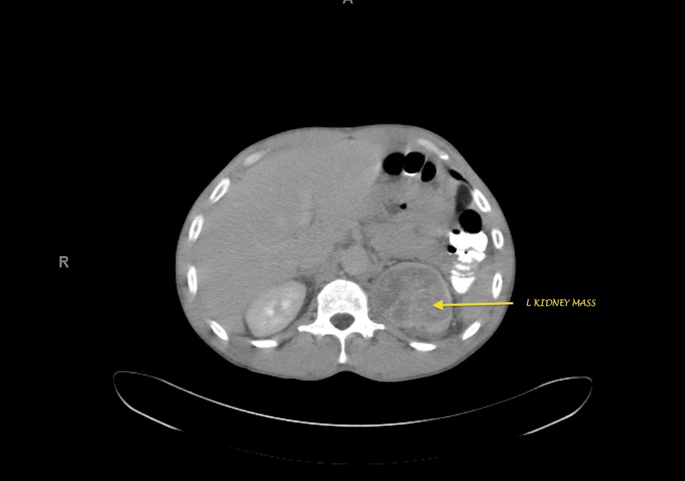
On this image, you see an abnormal interphase between the spleen and kidney. This could represent renal cell carcinoma or another invasive/metastatic process. Note the abnormal amorphous structure to the superior aspect of the L kidney. We do not see a clear border of the kidney and the overall structure is concerning for a mass. There is no appreciated free fluid. A CT scan of abdomen was obtained which further confirmed the US findings – see below image.

This patient was admitted for further work-up and management and is currently awaiting tissue confirmation of what is speculated to be RCC per hospital consultant notes.
2011

