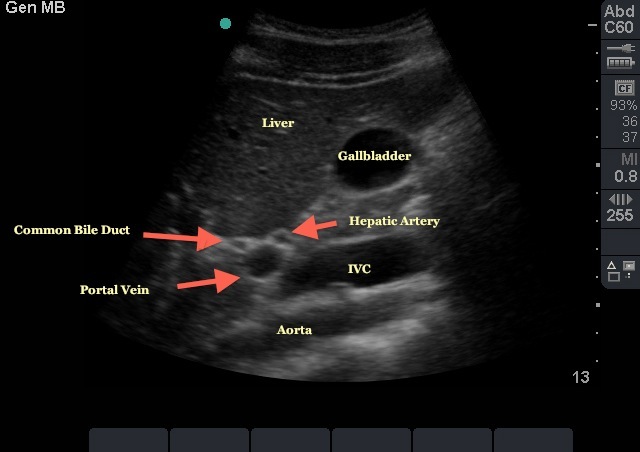
It is a short axis, or "Mickey Mouse," view of the portal triad obtained as part of a biliary scan. Once the gallbladder is visualized in its long axis this view can be obtained by following the main lobar fissure, which has the appearance of a thin hyperechoic line extending from the neck of the gallbladder to the portal vein. The portal vein appears as the dot at the base "exclamation point" formed in this longitudinal view. With subtle fanning and adjustments of the probe, the remaining structures of the portal triad (the hepatic artery and common bile duct) can be brought into view. Given the smaller size of these structures they often have the appearance of Mickey Mouse ears. If the probe indicator is towards the patient's right side, then Mickey's right ear should be the common bile duct. Given the absence of flow in the common bile duct, doppler imaging can be helpful to differentiate it from the portal vein and hepatic artery. Once this view is obtained the probe can be rotated 90 degrees to obtain a long axis view of the CBD for measurement. Measurement should be taken from inner wall to inner wall of the CBD. Normal width of the CBD is 4 mm for patients less than 50 with one additional mm allowed for every decade over 40.

2011

