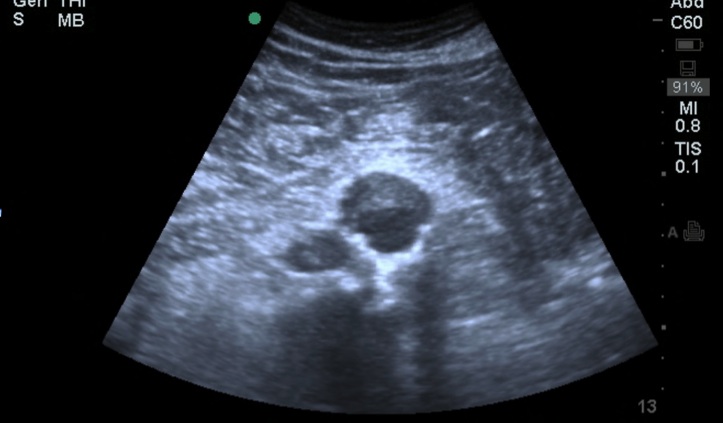
This week’s ultrasound image is an abdominal aorta scan on a hypotensive 64-year-old male who presented with abdominal and back pain (Image 1).
Image 1

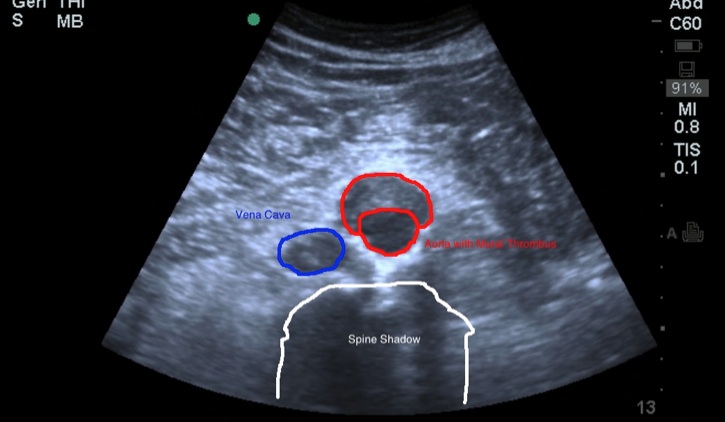
This image captures a transverse view of the abdominal aorta and raises concern for a luminal defect. Can you ID the anatomy and pathology? (Image 2)
Image 2

Remember these simple things when performing an aorta scan:
- The aorta is imaged using the curvilinear (abdominal) probe
- Gentle pressure on the probe will help disperse bowel gas and facilitate aortic views
- Obtain measurements of the aorta from outer wall to outer wall
- 5 images should be obtained for a full scan:
- Proximal aorta in cross section (indicator to the right)
- Mid Aorta in cross section (indicator to the right)
- Distal Aorta in cross section showing the bifurcation (indicator to the right)
- Iliacs showing individual diameter measurements (indicator to the right)
- One longitudinal view capturing the celiac trunk and SMA (indicator toward the head)

