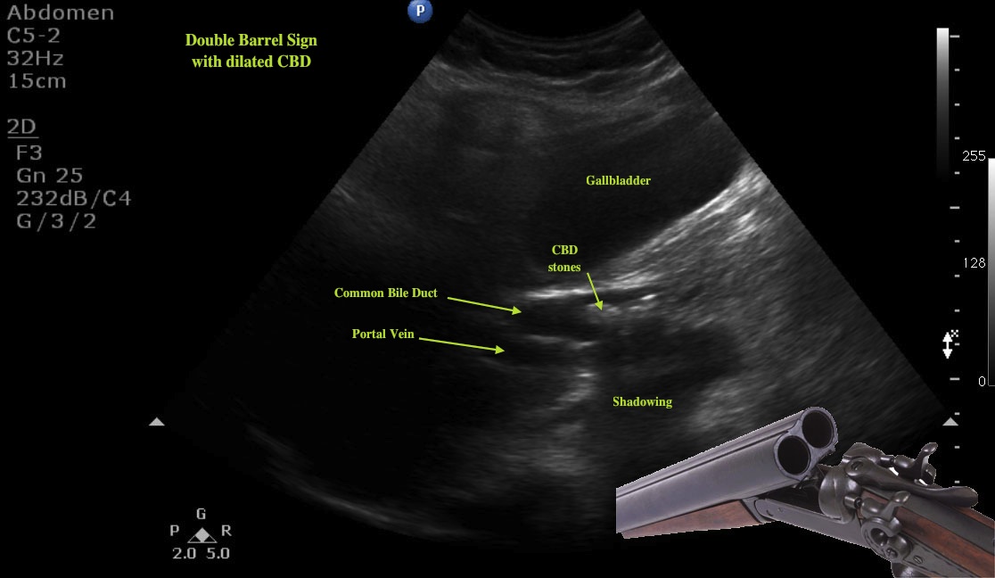
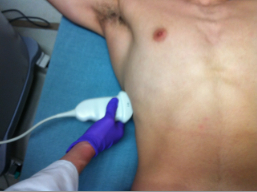
This week’s image shows us the “Double Barrel Shotgun” sign of a dilated common bile duct secondary to choledocholithiasis. Using the low frequency curvilinear probe with the indicator towards the patient’s head, the probe is swept from the xiphoid process out laterally beneath the costal margin until the gallbladder is visualized. We can obtain a “Mickey Mouse” view of the portal triad by locating the gall bladder in long axis, and then following the main lobar fissure to the portal vein in short axis. In this image we have found Mickey and then turn the probe 90 degrees so the portal vein and CBD are seen in long axis. The CBD runs just superior to the portal vein. The normal width of the CBD is 4mm for patients less than 50 with one additional mm allowed for every decade over 40. A normal CBD should be smaller than the portal vein beneath.
In this image the CBD is markedly dilated (1.3cm), roughly the same size as the portal vein, giving the appearance of a double barrel shotgun. Several small hyperechoic stones are seen within the CBD with associated inferior shadowing. These are not always seen and a dilated duct alone may be the only sign of CBD obstruction.


10/28/11

